

Resource > Scientific publications >
Exploring the potential of liver MPS of varied configurations to model cholestatic chemical effects
Filed under: DILI and Safety toxicology
Summary
Researchers used the PhysioMimix® MPS-LC12 liver-on-a-plate chip to test whether different human hepatocyte sources and culture formats can be used to detect the effects of cholestatic chemicals in vitro. Across primary human hepatocytes (PHHs), HepaRG cells, and induced pluripotent stem cell (iPSC)-derived hepatocytes (iHeps), the study found that PHH and HepaRG maintained comparable basal liver function in both 2D culture and the MPS-LC12, yet cholestasis-linked decreases in bile acid release were detected only in the MPS-LC12, with PHHs giving more consistent responses than HepaRG. The findings matter for toxicologists and drug safety teams because they show that a perfusion-based liver MPS can reveal cholestatic signals that static 2D models miss, supporting its use as a later-stage tool for liver injury assessment.
Study facts at a glance
| Publication | Nitsche KS, Sakolish C, Carmichael PL, Hewitt P, Bajaj P, Ferguson SS, Lloyd SM, Wilson SS, Bouwmeester H, Rusyn I. Exploring the potential of liver MOS of varied configurations to model cholestatic chemical effects. Archives of Toxicology. 2025. 100(3):1033-1047. |
| DOI | 10.1007/s00204-025-04263-1 |
| CN Bio product used | PhysioMimix® LC12 |
| How the platform was used | HepaRG cells and PHHs were cultured in the MPS-LC12 plates with recirculating media flow at 1 µL/s, seeded at approximately 600,000 cells per chip, for 7 days (both cell types) and up to 30 days (HepaRG), to measure basal liver function and responses to cholestatic chemicals. |
| Biological context | Human liver, DILI with a focus on cholestasis. Three hepatocyte sources were tested: HepaRG cells (a human hepatoma-derived cell line), single-donor PHHs, and iPSC-derived hepatocytes (iHeps). Healthy-source cells were exposed to known cholestatic chemicals. |
| Comparator | Conventional 2D static 96-well plate culture, plus two intermittent flow-based MPS (OrganoPlate 2-lane 96 and OrganoPlate 3-lane 40). Vehicle controls (0.1% DMSO) were used for chemical treatments. |
| Key readouts | Albumin secretion, urea secretion, CYP3A4 activity, LDH release, and total and individual bile acid concentrations (cholic acid, chenodeoxycholic acid, and their glycine and taurine conjugates) measured by LC-MS/MS. |
| Main interpretation | PHHs and HepaRG cells maintained comparable basal liver function in 2D culture and the MPS-LC12, but cholestasis-related decreases in bile acid release were detected only in the MPS-LC12, with PHHs giving more consistent responses than HepaRG. |
Table of Contents
- Summary
- Study facts at a glance
- Which CN Bio product was used?
- What this paper is about
- What the researchers found
- Why the paper matters
- Key study takeaways
- Why this paper is worth reading
- FAQs
- Full citation
- Related products and services
Which CN Bio product was used?
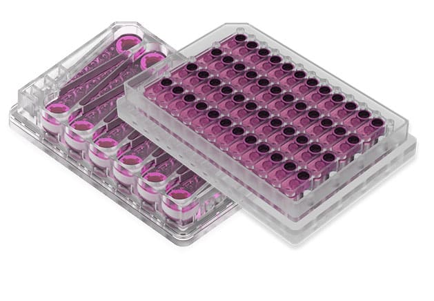
The study used the PhysioMimix MPS-LC12 plates, a constant perfusion-based liver MPS from. The PhysioMimix MPS-LC12 has the footprint of a standard microwell plate and contains 12 microfluidic chambers, each with a media reservoir and a culture well that holds a scaffold where cells are seeded. The flow was set at 1 µL/s. HepaRG cells and PHHs were seeded at approximately 600,000 cells per chip and cultured for 7 days, with one HepaRG experiment extended to 30 days. The MPS-LC12 was used to measure basal liver function (albumin secretion, urea secretion, and CYP3A4 activity) and, after treatment with cholestatic chemicals, to track changes in secreted bile acids. The two comparator MPS in the study, the OrganoPlate 2-lane 96 and 3-lane 40, were used to culture HepaRG and iPSC-derived hepatocytes. PHHs were not tested in the OrganoPlate models because they do not perform well in that format.
Find out more about CN Bio Liver-on-a-chip models here
What this paper is about
Drug-induced liver injury (DILI) is a leading cause of drug attrition and post-market withdrawal, and cholestatic and mixed injury types make up a sizable share of clinical cases. Cholestasis is difficult to reproduce in standard cell models, which limits early detection of compounds that disrupt bile acid handling. The paper addresses this gap by comparing how well three human hepatocyte sources, cultured across four formats, can report cholestatic effects. The cell sources were PHHs, HepaRG cells, and iPSC-derived hepatocytes. The formats were conventional 2D culture, the constant perfusion-based MPS-LC12, and two intermittent flow-based OrganoPlate models. The authors measured basal liver function over 7 days for most conditions and over 30 days for HepaRG in the MPS-LC12, then treated the cells with three compounds that cause cholestasis by different mechanisms: bosentan (BOS, 25 µM), alpha-naphthyl isothiocyanate (ANIT, 50 µM), and 2-octynoic acid (2-Oct, 70 µM). The aim was to identify which cell source and culture format combinations are suitable as in vitro models of cholestatic drug-induced liver injury.
Find out more about CN Bio DILI assays here
What the researchers found
For basal liver function, the study reported that PHHs and HepaRG cells performed at comparable levels in 2D culture and the MPS-LC12 over the first 7 days, with albumin higher in HepaRG and urea higher in PHHs. HepaRG function decreased in the second week, then recovered by weeks 3 and 4, and the cells maintained albumin, urea, and CYP3A4 activity for up to 30 days in the MPS-LC12. iPSC-derived hepatocytes showed low function in both 2D and OrganoPlate formats. In the OrganoPlate models, HepaRG produced more albumin and urea than iPSC-derived hepatocytes, but HepaRG function was lower than in 2D or the MPS-LC12.
For bile acid production, the study showed that after 7 days the total bile acid level was lowest in PHHs cultured in 2D and highest in PHHs cultured in the MPS-LC12, a 27-fold difference. HepaRG produced intermediate amounts, and the cell sources differed in their conjugation patterns: PHHs produced mainly glycine-conjugated bile acids, while HepaRG produced mainly taurine-conjugated bile acids.
For cholestatic compound effects, bosentan and ANIT produced marked CYP3A4 induction in PHHss and HepaRG in both 2D and the MPS-LC12, with the fold induction about twice as high in 2D as in the MPS. No CYP3A4 induction was seen in either OrganoPlate model, and 2-Oct did not induce CYP3A4. The key cholestasis readout was bile acid release: only in the MPS-LC12 did the compounds lower bile acid release into the medium. In PHHs, all three compounds caused a roughly twofold decrease, while in HepaRG only ANIT and 2-Oct had a significant effect at 7 days. In 30-day HepaRG cultures, only 2-Oct produced a significant decrease.
The findings within this publication demonstrate that not all liver models are equal for the same Context of Use (CoU), demonstrating the superior mechanistic and functional performance of PhysioMimix Liver-12 (LC12) plates in evaluating cholestatic drug effects that cause drug-induced liver injury (DILI) – a major challenge in drug safety assessment.
Why the paper matters
The study gives drug safety scientists and toxicologists practical guidance on choosing a liver model for cholestasis testing. It shows that a perfusion-based MPS can detect indirect-acting cholestatic compounds through changes in secreted bile acids, an effect that 2D culture did not capture. It also shows that 2D cultures can produce larger fold changes in CYP3A4 induction, which makes them useful for higher-throughput screening of metabolic induction, while the MPS-LC12 adds value for confirming findings and detecting cholestatic signals. Because secreted bile acids exceed intracellular bile acids and can be sampled from the larger medium volume of the MPS without disturbing the cells, medium sampling for bile acid analysis is a practical readout for longer studies. The work also supports HepaRG as an option where reduced donor variability and longer culture are needed, while noting that PHHs gave more consistent cholestatic responses. Taken together, the findings advance the use of in vitro liver models as part of NAMs for chemical and drug safety assessment.
Key study takeaways
- The study used the MPS-LC12 plate on the PhysioMimix platform to compare basal liver function and cholestatic responses across PHHs, HepaRG cells, and iPSC-derived hepatocytes.
- PHHs and HepaRG cells showed comparable basal function (albumin, urea, and CYP3A4 activity) in 2D culture and on the MPS-LC12 plate, while iPSC-derived hepatocytes performed poorly in all formats tested.
- Decreased bile acid release after cholestatic compound treatment was detected only on the MPS-LC12 plates, not in 2D culture or the OrganoPlate models.
- PHHs gave more consistent cholestatic responses than HepaRG: all three test compounds lowered bile acid release in PHHs, whereas HepaRG responded to a subset.
- CYP3A4 induction by bosentan and ANIT was about twice as high in 2D culture as onthe MPS-LC12 plates, indicating that 2D may suit higher-throughput induction screening.
- HepaRG maintained liver function for up to 30 days on the MPA-LC12 plates, supporting its use for longer-term, repeated-exposure studies where donor variability is a concern.
Why this paper is worth reading
This paper is useful because it provides side-by-side evidence for selecting a liver model based on the question being asked. A scientist deciding how to screen for cholestatic liability can see which cell source and culture format detected bile acid changes, which detected CYP3A4 induction, and where each format fell short. The data support a tiered approach: 2D culture with PHHs or HepaRG for concentration and time-course work, followed by the MPS-LC12 to confirm findings and capture cholestatic signals that depend on flow. For teams building safety packages under new approach methodologies, the study offers concrete readouts and culture conditions rather than general claims.
FAQs
The study used the MPS-LC12 liver plates on the PhysioMimix platform.
HepaRG cells and PHHs were cultured on the MPS-LC12 plates with a recirculating flow of 1 µL/s, seeded at about 600,000 cells per chip, for 7 days and up to 30 days for HepaRG, to measure basal liver function and bile acid responses to cholestatic chemicals.
The study modeled human cholestatic drug-induced liver injury using three hepatocyte sources: primary human hepatocytes, HepaRG cells, and iPSC-derived hepatocytes.
Cholestasis-related decreases in bile acid release were detected only in the PhysioMimix LC12, and primary human hepatocytes gave more consistent responses than HepaRG.
The study compared three hepatocyte sources across four culture formats: 2D culture, the PhysioMimix LC12, the OrganoPlate 2-lane 96, and the OrganoPlate 3-lane 40, under both basal conditions and treatment with bosentan, ANIT, and 2-octynoic acid.
The readouts were albumin secretion, urea secretion, CYP3A4 activity, lactate dehydrogenase release, and bile acid concentrations measured by LC-MS/MS.
The paper helps researchers choose a cell source and culture format for cholestasis testing, and it supports the use of the MPS-LC12 plates and the PhysioMimix platform as a flow-based model for detecting cholestatic signals that static 2D culture can miss.
Full citation
Related products and services
Contract research services
Discover how to utilize our cross-species models to inform next-step decision making via our DILI in vitro Contract Research Services here.
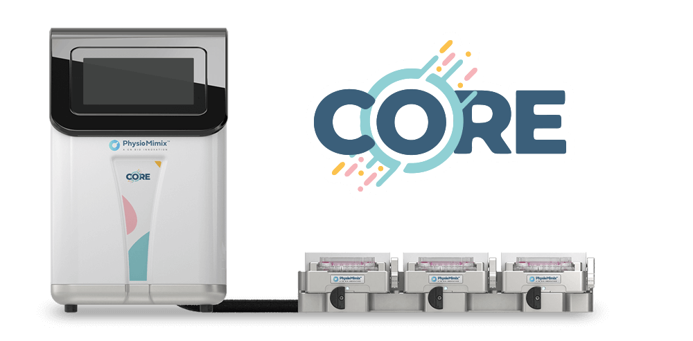
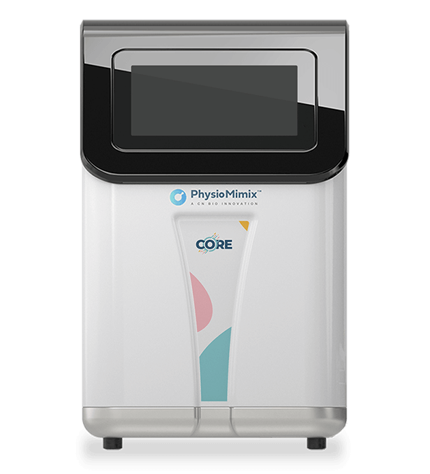
Add PhysioMimix Core in your lab
To develop your own cross-species Liver MPS models, you will need: