


Discuss your application with an expert
How can you predict immune-mediated liver injury following drug dosing?
Immune-mediated liver injury is caused by activation of inflammatory pathways, making the liver more sensitive to insults. In particular, new modality drugs, such as monoclonal antibodies, can activate the peripheral immune system to initiate a cascade of liver damage.
Immune-mediated liver injury is currently challenging to predict as the cause of the hepatic injury is often complex, interaction-based, and situation-specific. Therapeutics that act via the immune system, such as monoclonal antibodies for cancer, can under- or over-activate the immune system, causing increased autoimmunity or reactivation of liver viruses such as hepatitis B.
Traditional in vitro models do not facilitate the co-culture of liver cells with circulating immune cells, provide the longevity to explore chronic inflammation effects or enable the impact of underlying disease states on immune-mediated liver injury to be evaluated. In vivo models have unique immune systems which do not naturally recapitulate humans. In addition, stress from living in the laboratory setting and research can significantly impact metabolism, inflammatory responses and disease progressions.
Our solution
By incorporating immune cells into PhysioMimix® Liver-on-a-chip models, detailed mechanistic studies of the condition in the presence of various inflammatory cues and disease can now be performed to flag the immune-mediated liver injury risk of small molecules or biologics.
The tissue-resident macrophages of the liver (Kupffer cells) are cocultured with primary human hepatocytes to determine liver-specific inflammatory response as part of our PhysioMimix Drug-induced liver injury (DILI) assay.
Additionally, by including peripheral immune cells into the circulating media of the Liver-12 plate, it is possible to determine interactions between the immune system and the liver to identify adverse effects on hepatic health, function or changes to the inflammatory profile.
Using PhysioMimix disease models, drugs can also be tested against common liver disorders such as Hepatitis B and MASLD/MASH to identify patient cohorts with increased DILI susceptibility due to underlying inflammatory disease.
These capabilities enable you to future-proof your testing workflows in response to the FDA’s plans to phase out the animal testing requirement for monoclonal antibodies and other drugs with more human-relevant models. We are currently looking for partners to further validate the assay.
Please contact us if you would like to participate.

Poster
Recapitulating Immune-Driven Hepatotoxicity Using a Liver Microphysiological Platform

Studying immune-mediated liver injury
Limitations with current techniques
- Challenging to study immune-tissue level interaction in vitro
- Poor in vitro to in vivo data translation
- Unable to recapitulate inflammation driven by disease
- Challenging to run acute and chronic exposure assays due to short-lived cultures
Advancements with PhysioMimix Core
- Immune cells can be incorporated into and circulate around liver microtissues
- Measurement of clinical biomarkers (e.g. ALT/AST) allows for direct comparison to in vivo data
- Inflammation can be induced through disease state, e.g. MASLD/MASH
- Long-term stable tissues enable repeat compound dosing
End point measurements
Longitudinal and endpoint measurements include (but not limited to):
Functionality biomarkers
- Cytochrome P450 enzyme activity
- Albumin production
- Urea production
- Cytokine/chemokine release
Clinical liver health biomarkers
- Lactose dehydrogenase (LDH) release
- Adenosine Triphosphate (ATP)
- Aspartate Transferase (AST)/Alanine amino transferase (ALT)
Optional profiling analysis
- Quantitative PCR
- Transcriptomics
- Flow cytometry
Discern immune-mediated liver injury
Following exposure to pro-inflammatory stimuli, liver microtissues produce a range of physiologically relevant cytokines, chemokines, and growth factors that can be altered using different stimuli or inducing disease states.
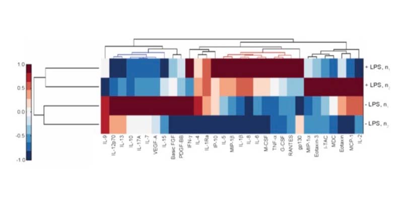
Flagging enhanced DILI risk due to inflammation
The effect of inflammation on DILI sensitivity is assessed by testing therapeutics with or without specific immunomodulators (e.g., LPS).


Detect immune-organ inflammatory events
Include peripheral immune cells and measure multiple soluble markers over time to understand interactions between multiple organs during inflammatory adverse events. Detect acute phase response proteins, like albumin and C-reactive protein (CRP), and predict cytokine storm events.


Frequently asked questions
1. To test for immune-mediated liver injury, should I use the DILI assay with or without peripheral blood cells?
This really depends on the question that you want to answer. If you are interested in detecting liver-based inflammation, our standard DILI assay is sufficient. Our DILI assay contains primary human hepatocytes and Kupffer cells, which are the resident macrophages of the liver. The presence of these cells enables the model to detect inflammatory initiation in the liver.
If you want to identify drug-induced activation of the adaptive immune system and the resulting interaction with the liver, peripheral immune cells can be added into the circulating flow of the chip (e.g., peripheral blood mononuclear cells (PBMCs) or isolated immune subtypes such as T-cells). We are currently looking for partners to further validate our immune-mediated liver injury assay. Please contact us if you would like to participate.
2. Why is CN Bio’s PhysioMimix Core System the best setup for investigating immune-mediated liver injury?
PhysioMimix Core technology features a closed-loop fluidic design which recirculates media around cultured microtissues, rather than single-pass fluidics. This approach facilitates concentration, rather than dilution of secreted biomarkers for easier detection. Plus, it enables the inclusion of circulating immune cells to understand their interactions with organs, the generation of an inflammatory response to stimuli or drugs, including monoclonal antibodies, and the study of inter-organ inflammatory crosstalk.
3. To what extent is donor matching of primary cells important when testing for immune-mediated liver injury?
It remains a challenge to donor match all cell types due to issues in sourcing all of the cell types from one donor. We have done some early work on HLA matching liver cells and peripheral immune cells, as have our customers. The outcome of these studies suggests that it is important to HLA-match to a certain degree. An SOT poster submitted by customers showed that, although PBMCs weren’t fully matched and some host-graft response was detected, the assay’s sensitivity was good enough to flag immune-mediated liver injury concerns found in the clinic.
4. Aside from the liver, which other CN Bio models can be used to study immune responses to drugs?
All PhysioMimix Multi-chip plates (Liver-12 &-48, Barrier-12 and Dual-Organ) use circulating flow, and therefore are conducive to the addition of circulating cells. Therefore, our Lung, lung/Liver and Gut/Liver models could be used for this purpose as well as the Liver. We have previously done proof-of-concept work using THP-1 monocyte cells in the Lung and Lung/Liver MPS models, whereby the monocytes were added to the circulating flow and various stimuli were used to test the inflammatory interactions between the immune cells, lung and liver microtissues. As of yet, we have not completed work in these models to test the inflammatory response to compounds; however, we know these models are immunocompetent and therefore will respond to an inflammatory challenge.
5. Are you planning to develop fully immunocompetent preclinical animal species DILI models?
Although these assays could potentially be useful for determining cross-species differences in drug responses, animal immune systems are very different from human and therefore, recapitulating these in vitro isn’t a high priority for us at the current time.
If you do not find the answer to your question listed, please contact us
Explore our Liver-on-a-chip models
Recreate the 3D multi-cellular architecture of the liver using perfused scaffolds. Achieve longer-term viability, enhanced functionality & high metabolic activity.
Access our DILI Service
Get instant access to the immune-mediated DILI Assay via our CRO Service. Through a collaborative approach, our experts work with you to plan and execute your study.
Bespoke projects are carried out by our dedicated team of scientists in our CRO facility, providing you with actionable data within weeks.
Add PhysioMimix Core to your lab
Harness the power of PhysioMimix Core in your own lab.
With a growing community of users and support from our experts, there has never been a better time to transition into 3D cell culture.