

Resource > Scientific publications >
IP-10 (CXCL10) Can Trigger Emergence of Dormant Breast Cancer Cells in a Metastatic Liver Microenvironment
Filed under: Disease modeling and Oncology
Summary
This study used the Legacy LiverChip, CN Bio’s perfused all-human liver microphysiological system (MPS), to test whether the chemokine IP-10 (CXCL10) can reawaken dormant triple-negative breast cancer (TNBC) cells in a liver metastatic niche. Dormant MDA-MB-231 cells surviving doxorubicin treatment within the hepatic niche emerged and outgrew in a dose-dependent manner when exposed to IP-10 at 0.5, 1.0, and 5.0 ng/mL, and the CXCR3 antagonist AMG-487 abrogated that outgrowth. The same concentrations applied to breast cancer cells alone in 2D proliferation, dormancy, migration, and invasion assays produced no significant change, identifying IP-10/CXCR3 as a candidate pathway for maintaining dormancy and showing why the cellular complexity of a perfused liver-on-a-chip model was needed to detect the effect at all.
Study facts at a glance
| Publication | Clark AM, Heusey HL, Griffith LG, Lauffenburger DA, Wells A. IP-10 (CXCL10) Can Trigger Emergence of Dormant Breast Cancer Cells in a Metastatic Liver Microenvironment. Frontiers in Oncology. May 27, 2021. 11:676135. |
| DOI | 10.3389/fonc.2021.676135 |
| CN Bio product used | PhysioMimix® Multi-chip Liver-12 plate and PhysioMimix Core System |
| How the platform was used | An all-human liver MPS was used to build a dormant-emergent metastatic niche: donor-matched primary human hepatocytes and non-parenchymal cells were seeded 1:1 onto collagen I coated scaffolds at 6 x 10⁵ cells per scaffold, triple-negative breast cancer (TNBC) cells were added on day 3, doxorubicin was applied on days 7 to 10 to leave only dormant cancer cells, and interferon gamma inducible protein-10 (IP-10, also known as CXCL10) was dosed on days 13 to 15 at 0.5, 1.0, or 5.0 ng/mL with or without the CXCR3 antagonist AMG-487 at 50 nM. Flow rate not specified. |
| Biological context | Liver metastatic niche in metastatic breast cancer. All-human hepatic tissue (donor-matched primary human hepatocytes plus non-parenchymal cells from 5 donors) colonized by RFP-labeled MDA-MB-231 TNBC cells, modeling dormancy and emergence rather than primary tumor growth. |
| Comparator | Within the liver MPS: doxorubicin-treated dormant cultures without IP-10, plus an AMG-487 inhibitor arm and a tumor-free hepatic niche. Outside the platform: conventional 2D assays (proliferation, Matrigel dormancy outgrowth, scratch migration, transwell invasion) in routine tumor medium (RPMI) and hepatocyte medium (HepM), and published human breast cancer expression and survival datasets. |
| Key readouts | Secreted CXCR3 ligand levels in MPS effluent (MIG/CXCL9, IP-10/CXCL10, I-TAC/CXCL11) by multiplex immunoassay; tumor burden as RFP+ scaffold area; cancer cell morphology as midpoint-to-length ratio; EdU incorporation, Matrigel outgrowth, scratch closure, and transwell invasion in 2D; RNA expression and survival in patients with stage IV breast cancer. |
| Main interpretation | IP-10 triggered dose-dependent emergence of dormant breast cancer cells within a perfused all-human hepatic niche, and the absence of a matching effect on breast cancer cells alone in 2D points to an indirect mechanism acting through the surrounding metastatic microenvironment. |
Table of Contents
Which CN Bio product was used?
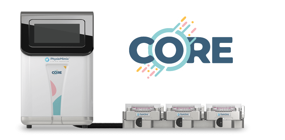
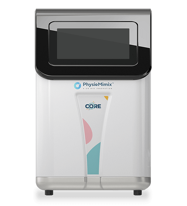
The study ran on CN Bio’s perfused 3D liver MPS, described in the paper under its former name, the Legacy LiverChip, and now supplied as the PhysioMimix Core Organ-on-a-chip (OOC) system. Each well holds a collagen-coated scaffold perfused with recirculating medium, which supports multi-cell-type hepatic tissue over the two-week culture period this protocol requires.
The metastatic niche was assembled directly in the platform. Donor-matched primary human hepatocytes and non-parenchymal cells were seeded at a 1:1 ratio, 6 x 10⁵ cells per scaffold, onto scaffolds coated with 1% rat-tail collagen type I in William’s E Medium with a thawing and plating supplement pack, then switched to maintenance medium after overnight culture. Once the hepatic tissue formed, 500 MDA-MB-231 cells expressing red fluorescent protein (RFP) were introduced on day 3, at which point a subpopulation spontaneously colonized the niche and adopted a dormant phenotype. Applicable cultures received 1 µM doxorubicin on days 7 to 10 to remove proliferating cancer cells, leaving the dormant population. They were stimulated on days 13 to 15 with IP-10 at 0.5, 1.0, or 5.0 ng/mL, with or without 50 nM AMG-487. Scaffolds were removed and fixed on day 15. Cells came from 5 donors, with 2 to 5 donors per outgrowth condition and 4 to 6 donors for the secreted chemokine measurements. Donor background, the presence of breast cancer cells, and treatment did not affect hepatic niche function or health.
The platform was used for the dormancy and emergence work only. All mechanistic follow-up on breast cancer cells in isolation, covering proliferation, Matrigel dormancy outgrowth, scratch migration, and transwell invasion, was performed in conventional 2D culture, and the human expression and survival analyses were drawn from published patient datasets. Imaging and quantification of cancer burden were carried out on scaffolds taken from the platform. The declaration of interests records that two authors hold a patent on the Legacy LiverChip technology commercialized by CN Bio Innovations.
Find out more about CN Bio Liver-on-a-chip models here
Find out more about CN Bio DILI assays here
What this paper is about
Breast cancer cells can disseminate early, colonize distant organs, and then persist as dormant cells for years before emerging as clinically detectable metastases. The liver carries the worst prognosis of any metastatic site in breast cancer, and the signals that awaken dormant cells there are largely unmapped. Identifying those signals matters because they are the targets a preventive strategy would have to act on.
This study addresses a gap that is as experimental as it is biological. Dormancy and emergence depend on interactions between disseminated tumor cells and the resident cells of the secondary organ, so a model must include both, keep them viable for weeks, and allow a dormant state to be established and then challenged. Conventional 2D assays measure tumor cell behavior in isolation. They cannot reproduce this arrangement, which is why this work used a perfused, all-human liver microphysiological system built from donor-matched hepatocytes and non-parenchymal cells. The candidate signals came from earlier gut-liver MPS work showing that CXCR3 ligands are synergistically upregulated under pathophysiological systemic inflammation. Of the three ligands (MIG/CXCL9, IP-10/CXCL10, and I-TAC/CXCL11), all bind the same receptor, CXCR3, so the authors focused on the most abundant one, IP-10. The study then asks a direct question: does IP-10 trigger emergence of dormant breast cancer cells in the liver niche, and if so, does it act on the cancer cells themselves or on the microenvironment around them?
What the researchers found
The main finding was that IP-10 triggered the emergence of dormant breast cancer cells in the perfused hepatic niche in a dose-dependent manner. Exposing dormant MDA-MB-231 cells to IP-10 at 0.5, 1.0, and 5.0 ng/mL for 48 hours produced significant outgrowth, measured as RFP+ scaffold area, and adding the CXCR3 antagonist AMG-487 significantly abrogated that effect. The doses were anchored to levels measured in effluent from the emergent group (approximately 1 ng/mL), with one dose on either side.
Analysis of effluent from the liver MPS showed that CXCR3 ligands were elevated on day 15 in niches containing actively growing cancer cells (both growing and emergent). In contrast, niches containing dormant cells resembled the tumor-free hepatic niche. IP-10 was present at markedly higher absolute levels than MIG or I-TAC, rising 2.2-fold in growing and 1.5-fold in emergent niches, compared with reported increases of 1.5-fold and 1.4-fold for MIG and 1.4-fold for I-TAC.
The model also showed a morphological shift. Cancer cells stimulated with IP-10 appeared more elongated than controls, reflected in a reduced midpoint-to-length ratio, which the authors interpret as a possible partial reversion toward a more mesenchymal phenotype.
The 2D work returned negative results, and they carry the argument. IP-10 stimulation was not associated with a significant increase in EdU incorporation in either routine tumor medium or hepatocyte medium, and did not significantly affect outgrowth of cells embedded in Matrigel. In scratch assays, IP-10 produced only a slight increase in migration in RPMI with no observable difference across doses that differed up to 500-fold. Meanwhile, the positive controls (lipopolysaccharide with epidermal growth factor and vascular endothelial growth factor) drove significant migration by 8 hours. In transwell invasion assays, 10% fetal bovine serum promoted invasion, while IP-10 alone showed only a modest increase. The study reported that IP-10 at levels comparable to those in liver MPS did not alter the proliferative, migratory, or invasive properties of MDA-MB-231 cells in vitro.
Clinical datasets supported the platform observations. In patients with stage IV disease, IP-10 was the predominant CXCR3 ligand and high expression was associated with significantly lower survival. IP-10 expression rose significantly with increasing tumor grade and was highest in patients with TNBC, and lowest in estrogen receptor positive and estrogen/progesterone receptor positive disease.
Why the paper matters
The practical value here is a worked case in which model choice determined whether a biological effect was detectable. The same chemokine, at comparable concentrations, produced significant outgrowth inside a perfused multicellular hepatic niche and nothing measurable against breast cancer cells cultured alone. For teams selecting a model for metastasis or dormancy research, that contrast sets a concrete expectation: signals that act through the microenvironment will not register in assays that contain only the tumor cell.
It also delivers a candidate pathway with a matching pharmacological tool. The reversal of IP-10-driven outgrowth by AMG-487 shows the effect is CXCR3-dependent and that the axis is tractable to antagonism in a human tissue model, supporting IP-10/CXCR3 as a target for approaches aimed at keeping disseminated cells dormant rather than treating established metastases. Pairing the MPS result with human expression and survival data strengthens the case that the observed behavior reflects what happens in patients, particularly in TNBC.
The scope is narrow, and the paper makes that clear. The study establishes that IP-10 triggers emergence and that the mechanism is indirect, and it identifies hepatic stellate cells and Kupffer cells (macrophages) as candidate responders based on existing literature. Determining which cell type in the niche relays the signal lies outside this work. For disease modeling groups, that unresolved step is itself the useful part: it marks where a deconvolution experiment in a similarly complex model would add the most.
Key study takeaways
- The study used CN Bio’s Legacy LiverChip, a perfused all-human liver microphysiological system, to recreate dormant and emergent metastatic breast cancer within a donor-matched hepatic niche of primary hepatocytes and non-parenchymal cells.
- The model showed that IP-10 (CXCL10) triggered emergence of dormant MDA-MB-231 triple-negative breast cancer cells in a dose-dependent manner across 0.5, 1.0, and 5.0 ng/mL, accompanied by a more elongated cancer cell morphology.
- Compared with dormant cultures without IP-10 and the CXCR3 antagonist AMG-487, the outgrowth response was CXCR3-dependent, as AMG-487 significantly abrogated it.
- Compared with the perfused hepatic niche, conventional 2D assays of proliferation, Matrigel dormancy outgrowth, scratch migration, and transwell invasion showed no significant response to IP-10, which indicates an indirect mechanism operating through the metastatic microenvironment.
- The workflow combined multiplex measurement of secreted CXCR3 ligands in MPS effluent, imaging-based quantification of tumor burden and cell morphology, 2D functional assays, and human expression and survival datasets from patients with stage IV breast cancer.
- The findings support the use of biologically complex, perfused liver models for mechanistic studies of metastatic dormancy, and the paper is explicit that the responding cell type within the hepatic niche remains to be identified.
Why this paper is worth reading
This paper is useful because it documents both a mechanism and the model conditions required to see it. Researchers working on metastatic dormancy, tumor microenvironment signaling, or liver metastasis get a defined experimental sequence, covering niche formation, spontaneous dormancy, chemotherapy selection, and cytokine challenge with a receptor antagonist control, that can be adapted to other stimuli and other secondary sites. Teams weighing whether the added complexity of a perfused multicellular organ-on-a-chip model justifies the effort will find a clean answer in the side-by-side comparison, where the 2D assays and the hepatic niche gave opposite results for the same chemokine. For translational and oncology drug discovery groups, the combination of a dose-dependent emergence phenotype, a pharmacological reversal, and supporting patient expression and survival data makes IP-10/CXCR3 a concrete starting point for dormancy-maintaining strategies rather than a general observation about inflammation and cancer.
FAQ
The study used the Legacy LiverChip, CN Bio’s perfused 3D liver microphysiological system, credited in the paper to CNBio Innovations Ltd. The platform is now supplied as the PhysioMimix Core Organ-on-a-chip system.
The Legacy LiverChip was used to build an all-human metastatic liver niche: donor-matched primary human hepatocytes and non-parenchymal cells seeded 1:1 at 6 x 10⁵ cells per collagen I coated scaffold, RFP-labeled MDA-MB-231 cells added on day 3, 1 µM doxorubicin on days 7 to 10 to isolate dormant cancer cells, and IP-10 at 0.5, 1.0, or 5.0 ng/mL with or without 50 nM AMG-487 on days 13 to 15. The publication does not specify the flow rate.
The model was a liver metastatic niche for metastatic breast cancer, built from donor-matched primary human hepatocytes and non-parenchymal cells from 5 donors, colonized by the MDA-MB-231 triple-negative breast cancer (TNBC) cell line, and used to study dormancy and emergence rather than primary tumor biology.
IP-10 (CXCL10) triggered dose-dependent emergence and outgrowth of dormant breast cancer cells within the liver microphysiological system, and the CXCR3 antagonist AMG-487 abrogated that outgrowth. Because IP-10 did not significantly alter proliferation, migration, or invasion of MDA-MB-231 cells alone, the paper concludes that IP-10 acts indirectly through the surrounding metastatic microenvironment.
The study compared IP-10-stimulated dormant cultures with doxorubicin-treated dormant cultures without IP-10, an AMG-487 inhibitor arm, and the tumor-free hepatic niche; it also compared responses in the perfused liver microphysiological system with conventional 2D proliferation, Matrigel dormancy, scratch migration, and transwell invasion assays run in two culture media.
Readouts included secreted MIG (CXCL9), IP-10 (CXCL10), and I-TAC (CXCL11) in MPS effluent by multiplex immunoassay, tumor burden as RFP+ scaffold area, cancer cell midpoint-to-length ratio, EdU incorporation, Matrigel outgrowth area, scratch closure, transwell invasion, and RNA expression with survival analysis in patients with stage IV breast cancer.
The paper gives metastasis and dormancy researchers a validated workflow for establishing and challenging dormant breast cancer cells in a perfused all-human liver microphysiological system, plus evidence that IP-10/CXCR3 is a candidate targetable pathway for maintaining dormancy. It also documents a case where a microenvironment-mediated effect was detectable in a complex 3D perfused model and not in simpler 2D assays, which is directly relevant to model selection.
Full citation
Related products and services
Contract research services
Discover how to utilize our cross-species models to inform next-step decision making via our DILI in vitro Contract Research Services here.
Add PhysioMimix Core in your lab
To develop your own cross-species Liver MPS models, you will need: