

Resource > Scientific publications >
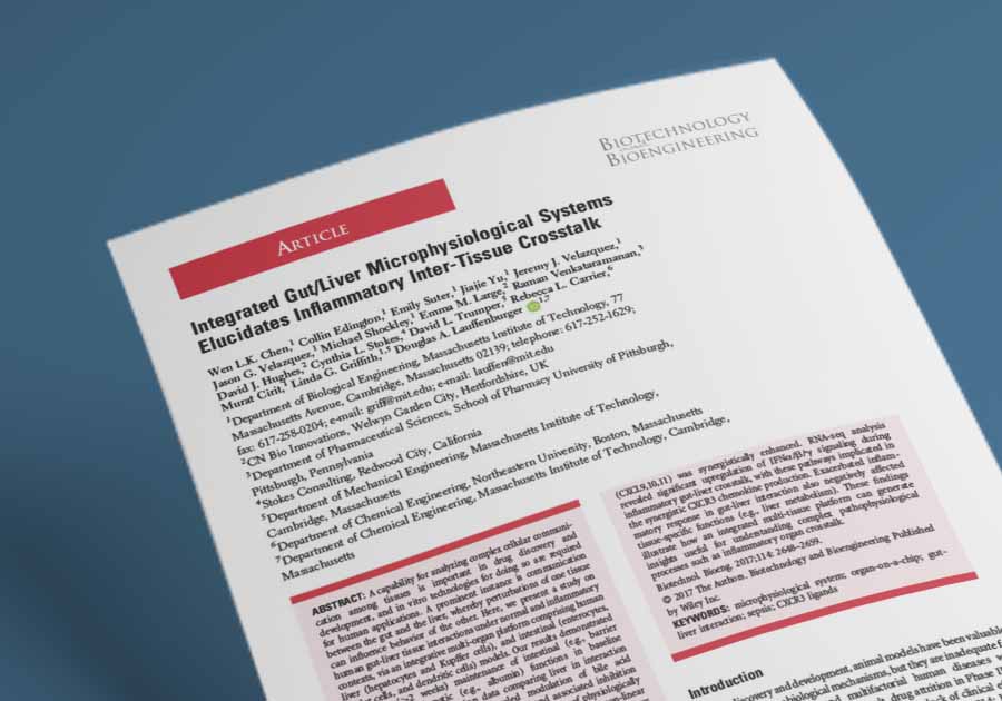
Integrated gut/liver microphysiological systems elucidates inflammatory inter-tissue crosstalk
Filed under: ADME, DILI, Disease modeling, Drug absorption, Drug metabolism, and Immune-mediated liver injury
Summary
Chen et al. (2017) fluidically linked an immune-competent human gut microphysiological system (MPS) with a human liver MPS on a multi-organ organ-on-a-chip platform built at MIT, using on-board pumping technology adapted from CN Bio’s LiverChip, the forerunner of PhysioMimix Core, to study gut-liver crosstalk under baseline and inflammatory conditions. The integrated gut-liver system maintained intestinal barrier function and liver albumin production for more than 2 weeks, showed suppression of the bile acid synthesis enzyme CYP7A1 alongside higher intestinal FGF19 secretion at baseline, and, under lipopolysaccharide (LPS)-induced inflammation, produced synergistic increases in CXCR3 chemokines together with broad suppression of hepatic cytochrome P450 (CYP) genes. The study, co-authored by CN Bio scientists, shows how a multi-organ MPS can expose inflammatory effects on liver metabolism that do not appear when each organ is cultured alone, which is relevant to disease modeling and to understanding drug metabolism during systemic inflammation.
Study facts at a glance
| Publication | Chen WLK, Edington C, Suter E, Yu J, Velazquez JJ, Velazquez JG, Shockley M, Large EM, Venkataramanan R, Hughes DJ, Stokes CL, Trumper DL, Carrier RL, Cirit M, Griffith LG, Lauffenburger DA. Integrated Gut/Liver Microphysiological Systems Elucidates Inflammatory Inter-Tissue Crosstalk. Biotechnology and Bioengineering. November 2017. 114(11):2648-2659. |
| DOI | 10.1002/bit.26370 |
| CN Bio product used | The study ran on a multi-microphysiological system (multi-MPS) platform built at the Massachusetts Institute of Technology (MIT), with on-board pneumatic pumping adapted from the technology used commercially in CN Bio’s LiverChip, the forerunner of the PhysioMimix® Core system. |
| How the platform was used | A polysulfone plate with on-board pumps linked a liver MPS, a gut MPS, and a mixing chamber at a systemic flow rate of 15 mL/day (75% of mixer outflow to the gut and 25% to the liver, with gut outflow feeding the liver as portal circulation), with internal recirculation of 1 µL/s in the liver and 0.25 µL/s in the gut, for up to 15 days of interaction. |
| Biological context | Human gut-liver axis under baseline conditions and simulated endotoxemia (lipopolysaccharide [LPS]-induced systemic inflammation). Liver: cryopreserved primary human hepatocytes and Kupffer cells at 10:1 on a perfused 3D scaffold. Gut: Caco-2 (C2BBe1) enterocytes and HT29-MTX goblet cells at 9:1 on Transwell® inserts, with primary human monocyte-derived dendritic cells on the basal side. |
| Comparator | Matched single-organ isolation controls on the same platform (liver with an empty gut compartment, and gut with an empty liver compartment) run with identical media volumes, flow rates, and flow partitioning; baseline versus LPS-stimulated conditions; and, for cytokines, the theoretical linear sum of the isolated gut and liver outputs. |
| Key readouts | Albumin secretion, transepithelial electrical resistance (TEER), mucin secretion, cytochrome P450 (CYP) activity by drug-substrate cocktail and mass spectrometry, RNA sequencing (RNA-seq) with Gene Ontology and gene set enrichment analysis, fibroblast growth factor 19 (FGF19) by enzyme-linked immunosorbent assay (ELISA), and secreted cytokines and chemokines at 6, 24, and 72 hours analyzed by hierarchical clustering, principal component analysis, and comparison against linear sums. |
| Main interpretation | Fluidically coupling immune-competent human gut and liver MPS preserved tissue-specific function for more than 2 weeks and revealed inter-organ effects absent from the single-organ controls, including FGF19-associated suppression of cholesterol 7α-hydroxylase (CYP7A1) at baseline, and non-additive production of C-X-C motif chemokine receptor 3 (CXCR3) ligands with broad suppression of hepatic CYP genes under inflammation. |
Table of Contents
Which CN Bio product was used?
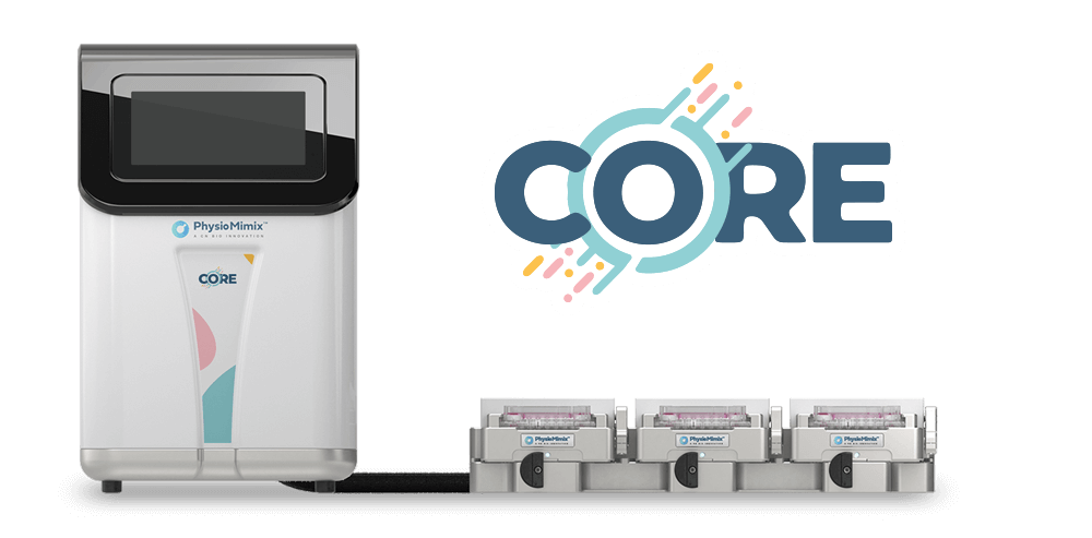
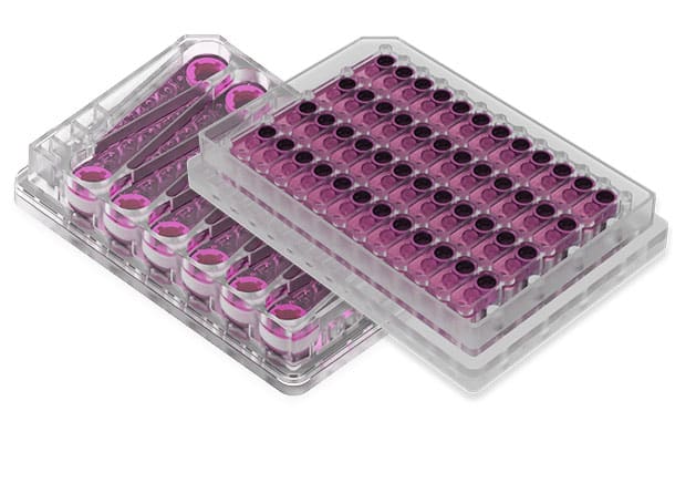
No commercial CN Bio product was used in this study. The gut-liver work ran on a multi-MPS platform built at MIT: a micromachined polysulfone top plate carrying the culture compartments and fluidic channels, a bottom plate that delivers compressed air and vacuum, and a polyurethane membrane between them that acts as the pump and valve layer, fastened into a single unit that is handled like a standard well plate. The on-board pneumatic pumping was adapted from technology used commercially in CN Bio’s LiverChip, and the liver compartment uses the same 301-channel perfused scaffold format.
The liver MPS contained 600,000 cryopreserved primary human hepatocytes and 60,000 Kupffer cells per well (10:1) on a collagen-coated polystyrene scaffold, seeded 3 days before interaction and recirculated at 1 µL/s in 1.6 mL of medium. The gut MPS combined Caco-2 (C2BBe1) and HT29-MTX cells at 9:1 on 12-well Transwell inserts, matured for 3 weeks off-platform, with monocyte-derived dendritic cells added to the basal side and a TEER acceptance threshold of at least 250 Ω·cm². The gut compartment held 0.5 mL apical and 1.5 mL basal medium, with the basal side recirculated at 0.25 µL/s, and the mixing chamber held 1.0 mL. Systemic flow of 15 mL/day was split 75% to the gut and 25% to the liver, and gut outflow fed the liver to represent portal circulation. All fluidically linked compartments shared a serum-free common medium containing 100 nM hydrocortisone, keeping cortisol in the physiological range. As a result, the liver retained its inflammatory responsiveness, and the medium was replaced every 3 days. Inflammation was induced with 2 ng/mL LPS in the circulating medium.
The Defense Advanced Research Projects Agency (DARPA) Microphysiological Systems Program and the National Institutes of Health (NIH) funded the study. CN Bio worked with MIT on a DARPA contract to develop a human body-on-chip system and, in 2019, secured an exclusive license to MIT’s multi-organ-on-chip technology from Linda Griffith’s laboratory to build multi-organ consumables for PhysioMimix Core. Researchers who want to run gut and liver co-culture on a commercial system can use CN Bio’s Gut/Liver-on-a-chip models, which pair a Caco-2/HT-29 or primary human jejunum gut barrier with primary human hepatocytes in the PhysioMimix Dual-organ plate.
Find out more about CN Bio Liver-on-a-chip models here
Find out more about CN Bio DILI assays here
What this paper is about
Communication between the gut and the liver shapes drug absorption, drug metabolism, and the body’s immune state. The liver receives most of its blood supply from the gut through the portal circulation, so it is continuously exposed to gut-derived metabolites, microbial products, and inflammatory mediators. The gut-liver axis also holds a large share of the body’s immune capacity: the gut is the largest immune organ, and the liver hosts over 70% of the body’s macrophages. Every preclinical model has strengths and limitations, and the authors note that interspecies differences often hinder predicting human responses from animal models, particularly for immune processes. That gap motivates human in vitro models that capture organ-to-organ communication.
Chen et al. built a dual-organ MPS with innate immune components in both tissues. The liver MPS co-cultured primary human hepatocytes with Kupffer cells, the liver’s resident macrophages, on a perfused scaffold similar to the one used in CN Bio’s Liver-on-a-chip models. The gut MPS combined absorptive enterocytes and mucus-producing goblet cells with primary monocyte-derived dendritic cells to represent key features of the small intestine.
The platform was machined from polysulfone rather than polydimethylsiloxane (PDMS), because hydrophobic compounds such as steroid hormones and many drugs partition into PDMS and prevent quantitative control of exposure. Each compartment has its own on-board pump, so medium recirculates within each tissue independently of the slower inter-organ exchange, the closed-loop recirculation principle that also underpins PhysioMimix organ-on-a-chip technology. Scaling choices were anchored to human physiology. The ratio of gut epithelial surface area to hepatocyte number is within the same order of magnitude as in vivo; Kupffer cells and hepatocytes are at 1:10, and dendritic cells and gut epithelial cells are at approximately 1:10. The study ran in parallel with a companion pharmacokinetic analysis in the same system, which is why gut surface area to hepatocyte number was chosen as the key scaling factor.
The researchers compared integrated gut-liver cultures with matched single-organ controls, first at baseline over 2 weeks and then under simulated endotoxemia, a state of elevated circulating LPS seen across clinical presentations including sepsis and inflammatory bowel disease (IBD).
What the researchers found
The main finding was that fluidically linked human gut and liver MPS behave differently from the same tissues cultured alone, and that the differences become much larger under inflammation.
At baseline, the study showed that hepatic and intestinal functions were maintained for 2 weeks and were comparable between interacting and isolated cultures. Albumin production agreed with levels previously reported for human hepatocyte cultures across multiple donors, TEER was comparable to published values for Caco-2/HT29-MTX co-cultures and primary intestinal monolayers, and mucin secretion was maintained. After 15 days, a CYP substrate cocktail showed that liver metabolic function was largely preserved, with CYP2C9 activity significantly higher and CYP3A4/5 activity lower in livers that had interacted with the gut.
RNA-seq after 3 days of baseline interaction identified 105 liver genes (70 upregulated, 35 downregulated) and 6 gut genes that changed significantly relative to isolation controls. Upregulated liver genes were enriched for cell division and cell cycle processes. Among the downregulated liver genes, CYP7A1, an enzyme central to bile acid synthesis, fell more than twofold, and the Gene Ontology terms linked to downregulated genes included bile acid biosynthesis and xenobiotic metabolism. However, these terms did not remain significant after multiple-testing adjustment. Intestinal FGF19, an established inhibitor of CYP7A1 in enterohepatic signaling, was higher in the interacting system at days 3 and 6, albeit at low concentrations, which the authors propose could explain the CYP7A1 suppression. The liver MPS has no separate biliary drainage. Still, hepatocytes release bile acids into the circulating medium, as shown in an earlier LiverChip study of an immune-competent hepatocyte-Kupffer cell co-culture, allowing bile acids to reach the gut’s basolateral side. The gut gene proprotein convertase subtilisin/kexin type 9 (PCSK9), which is involved in cholesterol homeostasis, was among the few intestinal genes that changed.
Adding 2 ng/mL LPS to the circulating medium for 3 days changed the expression of 2,548 liver genes (1,137 up, 1,411 down) and 780 gut genes (290 up, 490 down) in interacting versus isolated cultures. Both organs upregulated immune/defense response and interferon signaling pathways. The liver downregulated lipid and xenobiotic metabolism, and the gut downregulated alcohol biosynthesis and steroid and lipid metabolism. In the liver, expression of CYP1A2, CYP2C9, CYP2C19, CYP2D6, CYP3A4, and CYP3A5, enzymes that together metabolize over 90% of known drugs, was suppressed in the integrated system, which the authors suggest is likely due to accumulation of inflammatory mediators such as interleukin 6 (IL-6), tumor necrosis factor alpha (TNFα), and type I interferons.
Cytokine and chemokine profiling showed that inter-organ communication reshaped the inflammatory response. Principal component analysis of 72-hour data separated interacting from isolated cultures on the first component (76% of variance) and gut from liver on the second (20%). When measured levels in the integrated system were compared with the sum of the isolated gut and liver outputs, 58% of analytes were additive, 23% were less than additive, and 19% were synergistic. The strongest synergy involved the CXCR3 ligands CXCL10 (also known as interferon gamma-induced protein 10, IP-10) and CXCL11, with CXCL9 borderline significant. TNFα came predominantly from the gut, while both organs produced interferon gamma (IFNγ) at comparable levels.
To test the epithelium’s contribution, the researchers stimulated the gut epithelium alone, without immune cells, with 5 ng/mL TNFα, IFNγ, or both for 24 hours. Co-treatment markedly amplified 4 of the 8 synergistic chemokines seen in the integrated system (CXCL9, CXCL10, CXCL11, and CX3CL1). The study showed that TNFα and IFNγ crosstalk was central to the synergistic chemokine production and that intestinal epithelial cells contribute considerably to the overall immune milieu through paracrine interactions with immune cells.
Why the paper matters
For drug developers, the most direct implication concerns drug metabolism during inflammation. Patients with sepsis are prone to adverse drug reactions because inflammation suppresses hepatic CYP activity, and even modest CYP changes can cause toxicity for drugs with a narrow therapeutic window. This study showed that inflammatory gut-liver crosstalk suppressed expression of major drug-metabolizing CYP enzymes, leading the authors to conclude that predicting drug pharmacokinetics (PK) and pharmacodynamics (PD) requires attention to multi-organ interactions and disease context. A human gut-liver MPS provides a controlled setting to examine how systemic inflammation alters drug metabolism in the gut and in the liver.
In this model, the liver’s inflammatory response came from co-culturing primary human hepatocytes with Kupffer cells, the same pairing CN Bio uses to capture liver-specific inflammation in its work on immune-mediated liver injury. Chen et al. extend that principle across two organs, showing that the inflammatory output of a hepatocyte-Kupffer cell co-culture changes when the liver receives signals from an immune-competent gut.
The CXCR3 findings are relevant to disease modeling. CXCR3 signaling is implicated in autoimmunity, IBD, transplant rejection, infection, and cancer, and elevated CXCL9, CXCL10, and CXCL11 have been reported in the blood and intestinal biopsies of patients with IBD. By showing synergistic CXCR3 ligand production that arises only when the gut and liver communicate, the study positions gut-liver MPS as a controlled environment for mechanistic follow-up of inter-organ inflammation. The authors also report a recent finding that these CXCR3 ligands influence the emergence of dormant metastatic breast cancer cells in the liver MPS, and related work on IP-10 (CXCL10) and breast cancer dormancy in a metastatic liver MPS builds on this line of research.
The paper also sets out practical design choices for multi-organ MPS: PDMS-free materials for quantitative drug exposure, independent recirculation within each organ, physiological flow partitioning, and scaling by gut surface area to hepatocyte number. The authors report that cytokine concentrations on the platform were at the high end of ranges measured in patients with inflammatory conditions, occurring without plasma proteins, proteases, circulating immune cells, or other tissues that sequester, degrade, or take up cytokines in vivo. They note that directly scaling medium volume to cell number is not appropriate when cell types that produce or clear these factors are missing, and point to functional scaling as an emerging way to account for these differences.
Key study takeaways
- The study used an MIT-built multi-organ MPS platform, with pumping technology adapted from CN Bio’s LiverChip, to link an immune-competent human gut MPS with a liver MPS of primary human hepatocytes and Kupffer cells.
- At baseline, gut-liver interaction was associated with suppression of hepatic CYP7A1 and higher intestinal FGF19 secretion, consistent with known enterohepatic control of bile acid synthesis.
- Compared with the linear sum of isolated gut and liver cultures, 19% of measured cytokines and chemokines were synergistic under LPS stimulation, led by the CXCR3 ligands CXCL10 and CXCL11.
- The workflow combined 2-week functional markers (albumin, TEER, mucin, and CYP activity) with RNA-seq and time-resolved cytokine and chemokine profiling at 6, 24, and 72 hours.
- Inflammatory gut-liver crosstalk suppressed expression of the six CYP enzymes that together metabolize over 90% of known drugs, supporting the use of multi-organ MPS to study inflammation-mediated changes in drug disposition.
- The model is a minimal representation of the gut-liver axis: the gut uses cell lines, the liver lacks stellate and liver sinusoidal endothelial cells, adaptive immunity is not captured, immune cell migration was not tracked, and cytokines accumulate to the high end of patient ranges. The authors present it as a controlled environment for mechanistic investigation of inter-organ communication.
Why this paper is worth reading
This paper is useful because it pairs a fully documented multi-organ design, with flow partitioning, scaling ratios, media, and single-organ controls all specified, with a clear demonstration of what inter-organ communication adds: effects on bile acid regulation, cytokine output, and CYP expression that do not appear when the gut and liver are cultured separately. Teams planning multi-organ experiments can use its isolation-control design and linear-sum analysis as a template for separating direct tissue responses from crosstalk. Drug metabolism and toxicology scientists will find direct evidence that systemic inflammation can reshape hepatic CYP expression through the gut, and disease-modeling teams will find a worked example of tracing a synergistic chemokine response back to specific cytokines and cell types. For readers interested in the pharmacokinetic side of the gut-liver axis, a separate study applied a Caco-2/HT29 and primary hepatocyte gut/liver model on the PhysioMimix platform to the quantitative pharmacokinetics of the prodrug mycophenolate mofetil.
FAQ
Chen et al. (2017) did not use a commercial CN Bio product. The gut-liver MPS ran on a multi-organ platform built at MIT whose on-board pneumatic pumping was adapted from technology used commercially in CN Bio’s LiverChip, the forerunner of PhysioMimix Core, and CN Bio scientists Emma M. Large and David J. Hughes are co-authors.
The MIT multi-MPS platform linked a liver MPS, a gut MPS, and a mixing chamber at a systemic flow of 15 mL/day, sending 75% of mixer outflow to the gut and 25% to the liver, with gut outflow feeding the liver to mimic portal circulation. Each organ compartment had its own recirculation pump (1 µL/s for the liver and 0.25 µL/s for the gut), and cultures ran for up to 15 days at baseline or for 3 days with 2 ng/mL LPS.
The Chen et al. study modeled the human gut-liver axis under healthy baseline conditions and in simulated endotoxemia, a systemic inflammatory state linked to sepsis and inflammatory bowel disease. The liver MPS contained primary human hepatocytes and Kupffer cells, and the gut MPS contained Caco-2 enterocytes, HT29-MTX goblet cells, and primary human monocyte-derived dendritic cells.
The main finding was that fluidically linked human gut and liver MPS show inter-organ effects absent from single-organ cultures. These included FGF19-associated CYP7A1 suppression at baseline and, under LPS-induced inflammation, synergistic production of the CXCR3 ligands CXCL10 and CXCL11 with broad suppression of hepatic drug-metabolizing CYP genes.
Chen et al. compared integrated gut-liver cultures with gut-only and liver-only controls run on the same platform with identical media volumes and flow settings, under baseline and LPS-stimulated conditions. They also compared cytokine output from the integrated gut-liver system with the theoretical linear sum of the isolated gut and liver cultures to classify responses as additive, less than additive, or synergistic.
The gut-liver MPS study measured albumin, TEER, mucin, CYP activity by mass spectrometry, RNA-seq with Gene Ontology and gene set enrichment analysis, FGF19 by ELISA, and secreted cytokines and chemokines at 6, 24, and 72 hours.
The Chen et al. (2017) gut-liver MPS study shows how multi-organ organ-on-a-chip models can separate direct tissue responses from inter-organ crosstalk in human inflammation and drug metabolism research. It is most useful for disease modeling, drug metabolism, and toxicology teams studying how systemic inflammation alters liver function, given the model’s minimal cell composition and lack of adaptive immunity.
Full citation
Related products and services
Contract research services
Discover how to utilize our cross-species models to inform next-step decision making via our DILI in vitro Contract Research Services here.
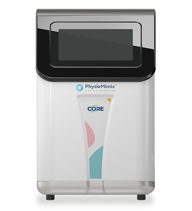
Add PhysioMimix Core in your lab
To develop your own cross-species Liver MPS models, you will need: