

Resource > Scientific publications >
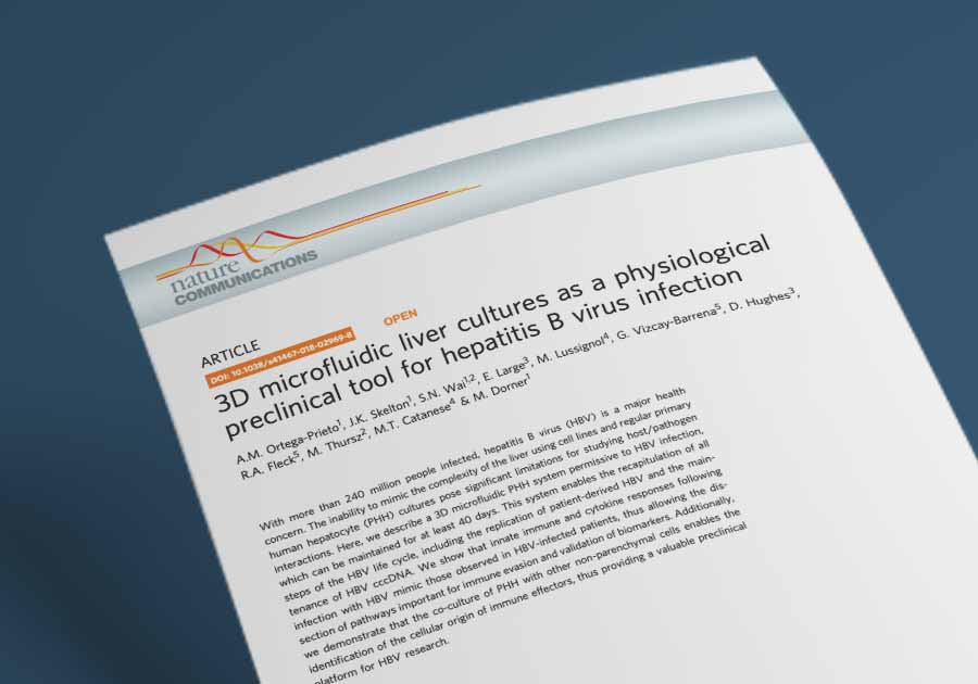
3D microfluidic liver cultures as a physiological preclinical tool for hepatitis B virus infection
Filed under: Disease modeling and HBV
Summary
Researchers at Imperial College London, King’s College London, and CN Bio Innovations used the CN Bio LiverChip perfused 3D liver microphysiological system to build a primary human hepatocyte model that is permissive to hepatitis B virus infection and stable for at least 40 days. The study reported that HBV established durable infection in the perfused 3D model at a multiplicity of infection (MOI) as low as 0.05 genome equivalents (GE) per cell, that infected microtissues accumulated cccDNA at approximately two copies per cell, and that infection suppressed type I and type III interferon responses in a pattern consistent with observations in patients. For drug discovery and infectious disease research, the model provides a human liver organ-on-a-chip system in which patient-derived HBV isolates, sequential antiviral regimens, and hepatocyte to Kupffer cell interactions can be studied in the same culture over weeks rather than days.
Study facts at a glance
| Publication | Ortega-Prieto AM, Skelton JK, Wai SN, Large E, Lussignol M, Vizcay-Barrena G, Hughes D, Fleck RA, Thursz M, Catanese MT, Dorner M. 3D microfluidic liver cultures as a physiological preclinical tool for hepatitis B virus infection. Nature Communications. Published online 14 February 2018; 9:682. |
| DOI | 10.1038/s41467-018-02969-8 |
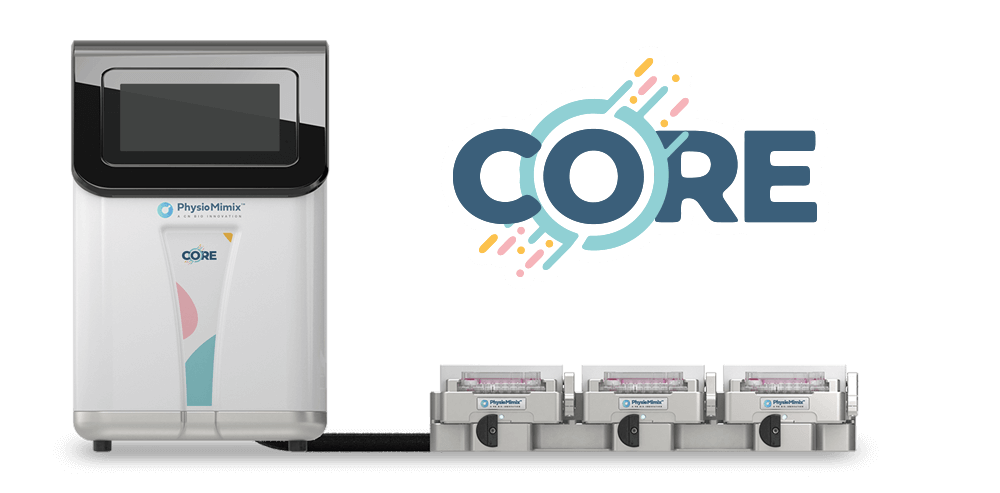
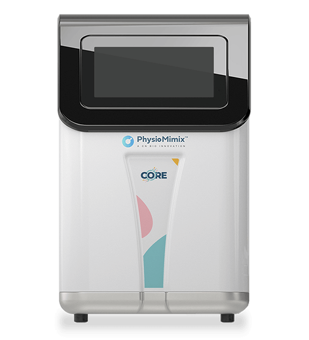
| CN Bio product used | PhysioMimix® Multi-chip Liver-12 plate and PhysioMimix Core System |
| How the platform was used | Cryopreserved primary human hepatocytes (PHH) were seeded at 600,000 viable cells per well in 1.6 mL of medium onto collagen-coated scaffolds, attached under 8 h of downward flow, then maintained under continuous upward recirculation at 1.0 µL/s for up to 40 days, as monocultures or as 10:1 co-cultures with primary human Kupffer cells (KC), and infected with cell culture-derived or patient-derived hepatitis B virus (HBV). |
| Biological context | Human liver, hepatitis B. Primary human hepatocytes from multiple donors, with primary human Kupffer cells as the non-parenchymal cell population. Infection performed in vitro with HepDE19-derived HBV and with HBV from the serum of five infected patients, benchmarked against serum cytokine levels in HBV-infected patients and healthy controls. |
| Comparator | Static 2D PHH cultures, 3D hepatic spheroids, self-assembling co-cultures of PHH with NIH3T3-J2 murine fibroblasts (SACC PHH), freshly thawed PHH, and HepG2 cells. Cytokine data were additionally compared with patient and healthy control sera. |
| Key readouts | Albumin secretion, lactate dehydrogenase (LDH) release, viability staining, cytochrome P450 (CYP450) gene expression, CYP1A2, CYP2C9, CYP3A4 and CYP3A activity, bile canalicular markers (dipeptidyl peptidase IV/CD26, 5-CDF staining), tight junction and polarization markers, transmission electron microscopy, hepatitis B surface antigen (HBsAg), hepatitis B e antigen (HBeAg) and hepatitis B core antigen (HBcAg), HBV DNA, covalently closed circular DNA (cccDNA), pregenomic and subgenomic HBV RNA, interferon (IFN) and interferon-stimulated gene (ISG) transcripts, pattern-recognition receptor expression, phospho-kinase arrays, and 102-plex cytokine and chemokine profiling with Luminex validation. |
| Main interpretation | Perfused 3D primary human hepatocyte cultures supported HBV infection at inocula several orders of magnitude below those required by static and spheroid models, sustained the full viral life cycle including cccDNA for at least 22 days, and reproduced the muted innate immune signature and several serum cytokine changes seen in HBV-infected patients, giving virologists a long-lived human liver model for host-pathogen and antiviral studies. |
Table of Contents
Which CN Bio product was used?
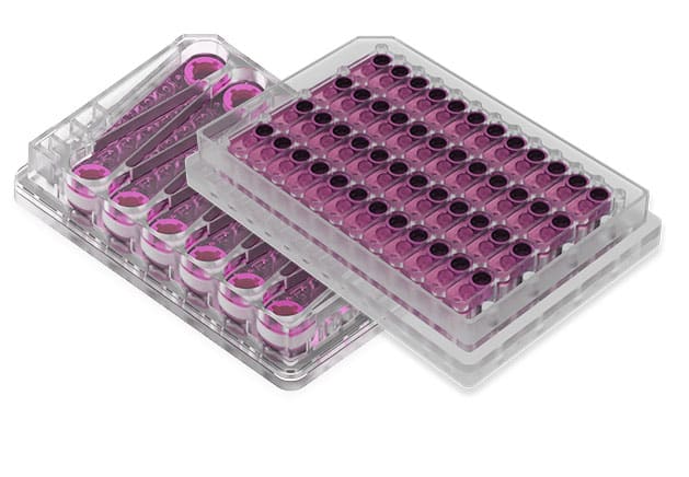
The study used the CN Bio LiverChip platform, a perfused MPS in which culture medium is recirculated by a pneumatically driven micropump through collagen-coated polystyrene scaffolds. Each plate holds 12 individually addressable bioreactors, the same 12-well perfused format supplied today as the Multi-chip Liver-12 plate, and flow was set at 1.0 µL/s: downward for the first 8 h to allow hepatocyte attachment within the scaffold, then upward for the remainder of the culture. Hepatocyte monocultures were seeded at 600,000 viable cells per well in 1.6 mL of medium, with medium replaced every 48 h, and PHH/KC co-cultures were seeded simultaneously at a 10:1 ratio. This is the technology CN Bio now supplies as the PhysioMimix Liver-on-a-chip model, run on the PhysioMimix Core system.
The platform was central to the 3D perfused arm of the study only. Comparator conditions were run outside it: 2D PHH in collagen-coated well plates, hepatic spheroids in ultra-low attachment 96-well plates, and SACC PHH in collagen-coated 96-well plates with murine fibroblast feeder cells. Analytical work including mass spectrometry, Luminex, cytokine arrays, quantitative PCR, and electron microscopy was performed on samples and scaffolds recovered from those cultures.
Two of the authors were affiliated with CN Bio Innovations at the time of publication, and the study was supported in part by funding from CN Bio Innovations alongside a Wellcome Trust New Investigator award, a European Research Council starting grant, and the Imperial NIHR Biomedical Research Centre.
Find out more about CN Bio Liver-on-a-chip models here
Find out more about CN Bio DILI assays here
What this paper is about
Chronic hepatitis B affects a very large global population, and the authors cite estimates of more than 240 million people infected. Progress in HBV research has been constrained by the models available. Hepatoma cell lines lose or alter expression of key innate immune sensors, and conventional 2D primary human hepatocyte cultures dedifferentiate within days, losing CYP450 activity and hepatocyte morphology before longer infection experiments can be completed. Most established systems also require very high viral inocula, sometimes up to 4 × 10⁴ GE per cell, together with dimethyl sulfoxide, polyethylene glycol, or inhibitors of innate immune signaling in order to detect infection. That combination sits awkwardly with what is known of HBV transmission in vivo, where small inocula are sufficient and no immunomodulation is involved.
The paper addresses that gap by testing whether restoring liver-like tissue architecture changes hepatocyte susceptibility to HBV. Primary human hepatocytes were cultured in a perfused 3D scaffold to generate polarized hepatic microtissues, then infected with cell culture-derived and patient-derived HBV and compared head to head with 2D cultures, 3D spheroids, and self-assembling co-cultures. The work spans model characterization, viral life cycle analysis, innate immune profiling, and co-culture with Kupffer cells to identify the cellular origin of individual immune effectors. Study areas covered include liver disease modeling, host-pathogen interaction, biomarker identification, and antiviral drug evaluation, each of which needs hepatocytes that stay differentiated for longer than a static culture allows, which is the problem perfused organ-on-a-chip technology is designed to address.
What the researchers found
The perfused 3D cultures formed stable, polarized hepatic microtissues. Primary human hepatocytes retained viability, morphology, and phenotype for at least 40 days, whereas 2D PHH and SACC PHH showed morphological dedifferentiation over 10 to 13 days. After two weeks of culture, the 3D perfused microtissues secreted approximately 10-fold more albumin per cell than 2D or SACC PHH, with albumin and LDH levels comparable to 3D spheroids. CYP450 gene expression in 3D perfused cultures and spheroids was comparable to freshly thawed PHH, while 2D cultures showed lower or absent expression. CYP3A activity was uniform across six hepatocyte donors at day 7, and CYP1A2, CYP2C9, and CYP3A4 activities were maintained at days 7, 14, and 21. Transmission electron microscopy at day 20 revealed bile canaliculi and tight junctions, and tight junction and hepatocyte markers (ZO-1, CD81, ITGB1, connexin 32) were present at day 26.
Infection was established at inocula orders of magnitude below those needed by other advanced models. Only the perfused 3D cultures remained infectable at 0.05 GE per cell, confirmed by HBV DNA secretion, although HBsAg at that inoculum fell below the limit of quantification. Susceptibility in 3D spheroids and SACC PHH was lost below 500 GE per cell. The authors reported this as infection at a 10,000-fold lower MOI than the other advanced culture models tested. Infection required neither polyethylene glycol, dimethyl sulfoxide, nor suppression of innate immune signaling, and was independent of hepatocyte donor and of the source of patient-derived virus.
The model supported the complete viral life cycle. Entry remained dependent on sodium taurocholate cotransporting polypeptide, since pretreatment with a myristoylated preS1 peptide inhibited HBsAg secretion by 90%, and heparinase III treatment abolished susceptibility altogether. Infected cultures accumulated pregenomic and subgenomic HBV RNA and maintained cccDNA at approximately two copies per cell, a level the authors note is similar to figures previously reported from human liver biopsies. Recombinant IFNα at 1000 IU/mL reduced HBV DNA and HBsAg secretion but left cccDNA levels largely unaffected, and tenofovir alafenamide reduced HBV DNA secretion while antigen expression remained stable.
Longevity enabled sequential treatment studies. Sequential IFNα followed by tenofovir alafenamide suppressed HBeAg and produced a more pronounced HBV DNA decline than either monotherapy. The p300 inhibitor C646 reduced secreted HBeAg and intracellular cccDNA, while IFNα monotherapy controlled HBV DNA release without the same effect on cccDNA.
HBV actively suppressed hepatocyte-intrinsic innate immunity. Type I and type III interferon transcripts in infected cultures fell to 10% of uninfected levels, with no baseline ISG induction. Expression of Toll-like receptors 1, 2, and 8, RIG-I, and IRF3 was reduced at 10 days post-infection and restored by IFNα treatment. Phospho-kinase arrays showed that a myristoylated preS1 peptide activated p38, JNK1/2/3, ERK1/2, and STAT2, whereas productive infection induced only ERK1/2 and overrode preS1-driven activation. Ultraviolet inactivation of the virus abolished the block, while heat denaturation did not restore immune activation, indicating dependence on conformational integrity of viral antigen.
Cytokine responses partially matched patient sera. Interleukin-8, MIP-3α/CCL20, Serpin E1, and MCP-1/CCL2 were significantly elevated both in infected 3D cultures and in the sera of HBV-infected patients. Vascular endothelial growth factor rose in culture but not in patient sera, and CXCL10 levels in culture were far lower than in patients, consistent with suppressed ISG induction in infected hepatocytes.
Kupffer cells required a secondary stimulus to respond. In 10:1 hepatocyte to Kupffer cell co-cultures, Kupffer cells remained viable and functional for at least 13 days and mounted an acute phase response, measurable as C-reactive protein, following infection. HBsAg, HBeAg, pregenomic RNA, and subgenomic RNA levels were nonetheless identical to monocultures. Kupffer cells secreted interleukin-6 and tumor necrosis factor α only after lipopolysaccharide stimulation, and that stimulation suppressed HBV replication as measured by reduced HBsAg secretion.
A stated limitation: no viral spread to neighboring uninfected cells was observed at low MOI, matching behavior in the other culture systems tested, which the authors interpret as evidence that additional host factors or cell populations contribute to spread through the liver.
Why the paper matters
Every preclinical model carries strengths and limitations, and HBV research has long had to work around the specific limitations of the systems available: short hepatocyte lifespan in 2D, altered innate immune sensor expression in cell lines, and inoculum requirements far above anything seen in transmission. This study addresses those constraints directly. By maintaining metabolically competent, polarized primary human hepatocytes under perfusion for weeks, the model allows infection with clinically relevant, patient-derived virus at low inoculum, and it keeps the culture alive long enough to run sequential dosing schedules of the kind used in combination trials.
Three points are practically useful for drug developers and translational teams. First, the model retains expression of innate immune sensors, so host response can be measured rather than inferred, and the suppression HBV exerts on that response becomes an experimental readout. Second, susceptibility across all tested donors opens the door to comparative work on host genetic background, including questions such as the contribution of IL-28B genotype to infection outcome. Third, the addition of Kupffer cells shows how the cellular origin of individual biomarkers can be assigned, something that is difficult in hepatocyte-only systems and in in vivo xenograft models. The same co-culture principle is applied elsewhere in the CN Bio portfolio, where adding Kupffer cells to primary human hepatocyte cultures raises sensitivity to innate immune-mediated responses in drug-induced liver injury studies.
Key study takeaways
- The study used the CN Bio LiverChip perfused 3D liver MPS, now supplied as the PhysioMimix Liver-on-a-chip model, to culture primary human hepatocytes under recirculating flow at 1.0 µL/s for up to 40 days.
- The 3D perfused microtissues reproduced liver sinusoid-like architecture with functional bile canaliculi, tight junctions, and hepatocyte polarization, alongside stable albumin secretion and CYP450 expression and activity.
- Compared with 2D PHH cultures, 3D spheroids, and SACC PHH, only the perfused 3D model established infection at 0.05 GE per cell, and it did so without polyethylene glycol, dimethyl sulfoxide, or innate immune inhibitors.
- The workflow combined virological readouts (HBsAg, HBeAg, HBV DNA, cccDNA, pregenomic and subgenomic RNA) with innate immune profiling (interferon and ISG transcripts, pattern-recognition receptor expression, phospho-kinase arrays) and 102-plex cytokine and chemokine analysis validated by Luminex.
- The findings support the use of perfused 3D primary human hepatocyte models for patient-derived HBV infection, sequential antiviral regimen testing, biomarker validation, and dissection of hepatocyte to Kupffer cell interactions.
- Context of use: the model is best suited to questions about establishment, maintenance, and treatment of infection in polarized human hepatocytes. It did not reproduce cell-to-cell viral spread at low MOI, so questions about spread through the liver need additional or alternative approaches.
Why this paper is worth reading
This paper is useful because it gives virologists and translational scientists a concrete basis for deciding which human liver model to use for HBV work, backed by a direct four-way comparison rather than a single-system description. It sets out the inoculum requirement, culture duration, infection kinetics, cccDNA burden, and innate immune behavior of a perfused 3D primary human hepatocyte model, and it reports which patient serum cytokine changes the model reproduces and which it does not. For teams designing antiviral studies, the sequential IFNα and tenofovir alafenamide and epigenetic modulator experiments show what a multi-week liver MPS assay can support that a short-duration static assay cannot.
FAQ
The study used the PhysioMimix® Core system with the Multi-chip Liver-12 plate. The plate provides 12 perfused, scaffold-based liver chips.
Primary human hepatocytes were seeded at 600,000 viable cells per well onto collagen-coated scaffolds in the CN Bio LiverChip platform and cultured under continuous medium recirculation at 1.0 µL/s for up to 40 days. Cultures were run as hepatocyte monocultures or as 10:1 hepatocyte to Kupffer cell co-cultures, and were infected with HBV three days after seeding.
The disease area is hepatitis B, and the model is a 3D perfused primary human hepatocyte culture forming polarized hepatic microtissues, with primary human Kupffer cells added for co-culture experiments. Infection was performed with HepDE19 cell culture-derived HBV and with HBV isolated from the serum of infected patients.
The paper reports the design of the first systematic, category-level comparison of eight commercially available liver MPS for a defined regulatory context of use, together with the harmonization, blinding, and contracting lessons from running it. DILI prediction results from the eight platforms are to be reported in a separate publication.
The study compared the 3D perfused primary human hepatocyte model with static 2D PHH cultures, 3D hepatic spheroids, self-assembling co-cultures of PHH with NIH3T3-J2 murine fibroblasts, freshly thawed PHH, and HepG2 cells. Cytokine responses in infected 3D cultures were additionally compared with sera from HBV-infected patients and healthy controls.
Readouts included albumin secretion, LDH release, viability staining, CYP450 gene expression and CYP1A2, CYP2C9, CYP3A4 and CYP3A activity, bile canalicular and tight junction markers, transmission electron microscopy, HBsAg, HBeAg, HBcAg, HBV DNA, cccDNA, pregenomic and subgenomic HBV RNA, interferon and ISG transcripts, pattern-recognition receptor expression, phospho-kinase arrays, and 102-plex cytokine and chemokine profiling validated by Luminex.
The paper is useful because it quantifies what a perfused 3D primary human hepatocyte model adds for hepatitis B research: infection with patient-derived HBV at low inoculum, cccDNA maintenance, weeks-long culture that supports sequential antiviral dosing, and preserved innate immune sensing that allows host response to be measured directly. It also states the limits of the model, including the absence of viral spread to neighboring uninfected cells at low MOI.
Full citation
Related products and services
Contract research services
Discover how to utilize our cross-species models to inform next-step decision making via our DILI in vitro Contract Research Services here.
Add PhysioMimix Core in your lab
To develop your own cross-species Liver MPS models, you will need: