

Resource > Scientific publications >
Morphological behaviour and metabolic capacity of cryopreserved human primary hepatocytes cultivated in a perfused multiwell device
Filed under: ADME and Drug metabolism

Summary
Researchers at Sanofi R&D used CN Bio’s perfused multiwell liver device, named LiverChip in the paper, to culture cryopreserved primary human hepatocytes from three donors for 7 days and compare their drug-metabolizing capacity against the same hepatocyte preparations in 2D static culture. The perfused microtissues retained differentiated hepatocyte morphology, bile canaliculi, and rising albumin secretion across the week, and mean in vitro intrinsic clearance measured on Day 4 was 7.3-fold higher for phenacetin (CYP1A2), 6.4-fold higher for midazolam (CYP3A), and 2.4-fold higher for dextromethorphan (CYP2D6) than in Day 1 2D static culture, while bupropion (CYP2B6) and tolbutamide (CYP2C9) clearances were broadly comparable between the two formats. For DMPK and absorption, distribution, metabolism, and excretion (ADME) teams, the study provides donor-level evidence that a perfused liver microphysiological system (MPS) can sustain phase I and phase II metabolic function long enough to support incubations that outlast conventional hepatocyte monolayers.
Study facts at a glance
| Publication | Vivares A, Salle-Lefort S, Arabeyre-Fabre C, Ngo R, Penarier G, Bremond M, Moliner P, Gallas JF, Fabre G, Klieber S. Morphological behaviour and metabolic capacity of cryopreserved human primary hepatocytes cultivated in a perfused multiwell device. Xenobiotica. 2015;45(1):29-44. |
| DOI | 10.3109/00498254.2014.944612 |
| CN Bio product used | CN Bio’s perfused multiwell liver device, referred to throughout the paper by its former name, LiverChip. The plate contains 12 individually addressable bioreactors, each comprising a culture well with a collagen-coated scaffold, a reservoir, and an integrated micropump that recirculates medium through the scaffold. |
| How the platform was used | Cryopreserved primary human hepatocytes were seeded at 0.6×10⁶ cells per well in 1.4 mL, attached to the scaffold for 8 hours under downward flow at 1 µL/second, then cultured under recirculating perfusion for 7 days, with cytochrome P450 (CYP) probe substrate incubations run on Day 4 and midazolam kinetics repeated on Day 7. |
| Biological context | Healthy human liver. Cryopreserved primary human hepatocytes from three donor preparations (BD295, BD304, and QHu4019), cultured as a perfused three-dimensional (3D) liver microtissue monoculture with no non-parenchymal cells and no disease model. The application area is drug metabolism and pharmacokinetics (DMPK) rather than disease modeling. |
| Comparator | 2D static monolayer culture of the same hepatocyte preparations in collagen I coated 24-well microplates, assayed on Day 1 after seeding. Selective CYP inhibitors (furafylline, sulphaphenazole, quinidine, and ketoconazole) and the pan-CYP inhibitor 1-aminobenzotriazole (ABT) were used as mechanistic controls in both conditions. |
| Key readouts | Optical microscopy of microtissue formation; live/dead fluorescence staining; daily albumin secretion by enzyme-linked immunosorbent assay (ELISA) and albumin immunofluorescence; scanning electron microscopy (SEM) and transmission electron microscopy (TEM) ultrastructure; reverse transcription quantitative polymerase chain reaction (RT-qPCR) of 22 genes covering phase I enzymes, phase II enzymes, uptake and efflux transporters, and nuclear receptors; liquid chromatography with tandem mass spectrometry (LC-MS/MS) quantification of five CYP probe substrates and their metabolites; in vitro intrinsic clearance (CLint; non-specific binding of probe compounds to the device. |
| Main interpretation | Cryopreserved primary human hepatocytes maintained differentiated morphology, albumin secretion, and phase I and phase II drug-metabolizing activity for at least 7 days under perfusion, with CYP1A2, CYP3A, and CYP2D6 activities higher than in Day 1 2D static culture and CYP2B6 and CYP2C9 activities comparable, which extends the usable window for hepatic clearance work beyond that of conventional hepatocyte monolayers. |
Table of Contents
Which CN Bio product was used?
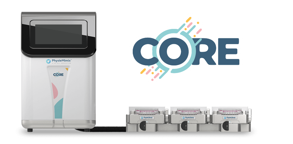
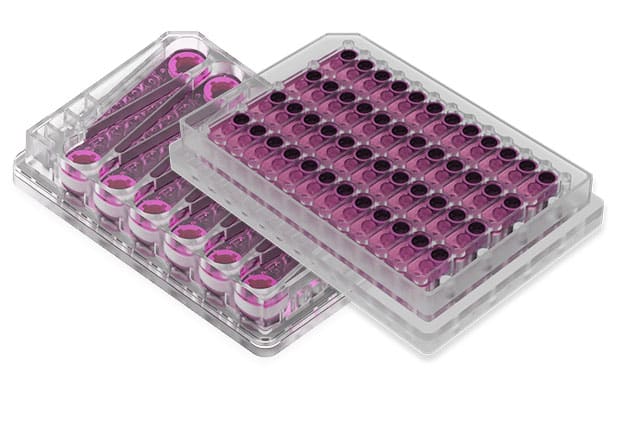
The study used CN Bio’s perfused multiwell liver device, described in the paper under its former name, LiverChip, and now supplied as the PhysioMimix® Organ-on-a-chip (OOC) systems with liver microtissues cultured in the Multi-chip Liver-12 plate. Each plate contains 12 isolated bioreactors, and each bioreactor holds a collagen-coated scaffold engineered to recapitulate the dimensions of the liver sinusoidal capillary bed, together with a reservoir and a pneumatically driven micropump that recirculates culture medium through the scaffold. Cryopreserved primary human hepatocytes were seeded at 0.6×10⁶ cells per well in 1.4 mL of seeding medium and allowed to attach to the scaffold for 8 hours at 37 °C under a downward flow of 1 µL/second, after which flow was reversed to pass upward through the scaffold. Medium was exchanged for long-term culture medium the following day, and after three further days of culture the hepatocytes were incubated with FDA-recommended CYP probe substrates. The platform was used only for the perfused arm of the study. The 2D static comparator was run in collagen I coated 24-well microplates at 0.42×10⁶ cells per well, and all substrate and metabolite quantification for both arms was performed off-platform by LC-MS/MS. Gene expression analysis, microscopy, and albumin measurements were likewise carried out on samples and scaffolds taken from the device. The work was designed and run by Sanofi R&D.
What this paper is about
Predicting human metabolic clearance from in vitro data is difficult for low-turnover compounds, because the assay has to run long enough for a measurable amount of the compound to disappear. Conventional primary hepatocyte monolayers do not offer that window: CYP-dependent activity falls substantially during the first 24 to 48 hours of static monolayer culture, and viability declines after around Day 3. The practical consequence for DMPK groups is that slowly metabolized compounds and longer-duration hepatotoxicity questions fall outside what a standard suspension or monolayer assay can address.
This paper asks whether continuous perfusion of a 3D hepatocyte microtissue holds that window open. The model is a monoculture of cryopreserved primary human hepatocytes seeded onto a collagen-coated scaffold inside a perfused bioreactor, cultured for 7 days, with three separate donor preparations used to give some sense of donor-to-donor spread. The comparison is a direct one against the format the field already relies on: the same three hepatocyte preparations in 2D static culture on collagen I, assayed on Day 1 after seeding, which the authors selected as the point at which static metabolic activity is highest and before global dedifferentiation sets in. Perfused cultures were assayed on Day 4, once the microtissues had formed. The paper covers three linked questions across that design: whether hepatocyte structure and viability hold up, whether mRNA expression of drug metabolism and transport genes is preserved, and whether measurable phase I and phase II activity is retained. It sits in the same body of end-user evidence as the organ-on-a-chip end user perspective from GSK, in that the work was run inside a pharmaceutical DMPK group on its own terms.
What the researchers found
The main finding was that metabolic capacity in the perfused device on Day 4 matched or exceeded that of Day 1 2D static culture across five probe substrates. Mean in vitro intrinsic clearance was 7.3-fold higher for phenacetin (0.277 versus 0.038 mL/h/106 cells), 6.4-fold higher for midazolam, and 2.4-fold higher for dextromethorphan under perfusion, while bupropion was 1.3-fold higher and tolbutamide was slightly lower at 0.7-fold. Measured as percentage biotransformation over a 24-hour incubation, phenacetin conversion to 4-acetamidophenol rose from a mean of 8.5% in static culture to 35.6% under perfusion, a 4.2-fold increase, with smaller increases for bupropion (1.9-fold), tolbutamide (1.3-fold), midazolam (1.3-fold), and dextromethorphan (1.1-fold). In the discussion the authors summarize this as roughly 7-fold and 6-fold higher CYP1A2 and CYP3A activity, around 2-fold higher CYP2D6 activity, and conserved CYP2B6 and CYP2C9 activity relative to 2D static culture.
The study also reported that CYP3A activity was stable across the culture period rather than merely elevated at one timepoint. Midazolam disappearance and metabolite formation were near-identical on Day 4 and Day 7 in one donor preparation, with intrinsic clearance of 1.04 mL/h/106 cells on Day 4 against 1.28 mL/h/106 cells on Day 7. On both days, more than 75% of the initial midazolam concentration was metabolized within 6 hours and approximately 90% within 24 hours.
Phase II conjugation was active alongside the CYP reactions. Formation of dextrorphan O-glucuronide was considerably higher under perfusion than in static culture, which the authors attribute to higher UDP-glucuronosyltransferase (UGT) 2B activity, and midazolam was conjugated both as 1′-hydroxy-midazolam glucuronide via the CYP3A and UGT2B4/7 pathway and as direct N-glucuronides via UGT1A4. Reaction phenotyping behaved as expected under flow: furafylline abolished 4-acetamidophenol formation, sulphaphenazole abolished tolbutamide metabolite formation, quinidine reduced dextrorphan and its glucuronide, ketoconazole suppressed the CYP3A-dependent hydroxyl metabolite of dextromethorphan and shifted flux toward the CYP2D6 pathway, and ABT abolished CYP-dependent metabolite formation altogether.
At the gene expression level, RT-qPCR of 22 genes showed that basal CYP mRNA levels fell during the first two days of perfused culture and were then conserved from Day 2 to Day 7 for most genes. CYP2B6 and UGT1A6 were persistently up-regulated, the four UGT genes measured were unchanged or up-regulated after Day 1, the nuclear receptors PXR and CAR were unchanged or slightly down-regulated, and efflux transporter genes BCRP, MRP2, and MDR1 were maintained or moderately up-regulated. Against that, OATP1B3 was strongly reduced, BSEP was down-regulated, and CYP2C8, CYP2C9, and CYP2D6 declined over the same period. Because mRNA profiles differed markedly between donors, the authors report only tendencies and did not calculate standard deviations for the gene expression data.
Structural and functional readouts supported the metabolic data. Live/dead staining showed high viability with few dead cells after 7 days, and daily albumin secretion rose from Day 1 and plateaued by Days 5 to 7. SEM showed hepatocytes uniformly lining the scaffold channel walls as a layer approximately 80 to 100 µm thick. TEM showed features typical of viable hepatocytes in vivo: bile canaliculi with microvilli, cell junctions, abundant mitochondria, rough endoplasmic reticulum, glycogen storage, and occasional lipid inclusions. Both SEM and TEM also showed debris and necrotic hepatocytes expelled to the luminal surface, consistent with the small dead-cell population detected by ethidium homodimer-1 staining. A separate control experiment circulated the five probe compounds through the perfusion circuit and scaffold without cells, and found that less than 20% of each compound was bound to the device after 24 hours.
Why the paper matters
For DMPK groups choosing an assay format, the practical value here is the length of the usable window rather than any single fold-change. A static hepatocyte monolayer gives its best metabolic readout on Day 1 and deteriorates from there, which caps how slowly a compound can turn over before the assay runs out of viable tissue. This study shows measurable CYP1A2, CYP2B6, CYP2C9, CYP2D6, and CYP3A activity on Day 4 of perfused culture, and equivalent CYP3A activity again on Day 7, in tissue that still displays bile canaliculi and rising albumin output. That combination is what makes longer incubations feasible, and it is the reason the authors position the model for future work on low-clearance compounds and in vitro to in vivo extrapolation (IVIVE).
The paper is also useful for what it settles about assay mechanics. Non-specific binding to microfluidic devices is a recurring objection to perfused formats, and the authors addressed it directly with a cell-free circulation control before running any activity work. Selective inhibitor responses under flow mean that reaction phenotyping and fraction metabolized questions can be asked on the platform rather than only on the static comparator. Together these points make the device a candidate for the metabolism work that sits upstream of drug-induced liver injury (DILI) assessment, and they matter for teams building a case for human-relevant organ models in a regulated workflow.
Key study takeaways
- The study used CN Bio’s perfused multiwell liver device, named LiverChip in the paper, to culture cryopreserved primary human hepatocytes from three donors for 7 days under recirculating flow.
- The model retained a differentiated hepatocyte phenotype across the week, with bile canaliculi, microvilli, cell junctions, glycogen storage, high viability, and daily albumin secretion that rose from Day 1 and plateaued by Days 5 to 7.
- Compared with Day 1 2D static culture, mean in vitro intrinsic clearance on Day 4 under perfusion was 7.3-fold higher for phenacetin, 6.4-fold higher for midazolam, and 2.4-fold higher for dextromethorphan, with bupropion and tolbutamide broadly comparable.
- The workflow combined RT-qPCR of 22 drug metabolism and transport genes, LC-MS/MS substrate and metabolite quantification, intrinsic clearance calculation, selective and pan-CYP inhibitor controls, albumin ELISA, and SEM and TEM ultrastructural analysis.
- Phase II conjugation was retained under flow, with higher dextrorphan O-glucuronide formation indicating UGT2B activity and midazolam N-glucuronidation indicating UGT1A4 activity.
- The findings support the use of a perfused liver MPS for metabolism studies that need to run for several days, with the caveats that the model is a hepatocyte monoculture, gene expression data were reported as donor-level tendencies only, and the Day 4 to Day 7 clearance comparison rests on a single donor preparation.
Why this paper is worth reading
This paper is useful because it was run by a pharmaceutical DMPK group on its own hepatocyte preparations, against the static format that group already used, and it reports the unfavorable comparisons alongside the favorable ones. Anyone deciding whether a perfused liver model earns a place in a clearance workflow gets the specific numbers they need: which CYP activities gain under flow, which are merely preserved, how much compound binds to the device, whether selective inhibitors still behave predictably, and how far into the culture period CYP3A activity holds. It also gives a concrete operating protocol, from seeding density and attachment flow direction through to the day on which probe incubations were performed, which makes it a practical reference for scoping a study rather than only a demonstration of feasibility. Teams weighing that decision may also want to see how similar workflows are run as contract research services or documented as step-by-step MPS protocols.
FAQs
The study used CN Bio’s perfused multiwell liver device, which the paper calls LiverChip, comprising 12 individually addressable bioreactors, each with a collagen-coated scaffold, a reservoir, and an integrated micropump. CN Bio’s current equivalent platform is the PhysioMimix Organ-on-a-chip (OOC) systems with the Multi-chip Liver-12 plate.
The liver/immune MPS was used to measure HC pharmacokinetics over 48 hours at low (1.25 mg/mL) and high (25 mg/mL) HSA concentrations, sampled across culture days 3 to 5 and days 5 to 7, and to measure TNF-α and IL-6 secretion following LPS doses of 1 µg/mL repeated every 48 hours with complete media changes. The four-MPS interactome in the same paper was simulated computationally, not built on the platform.
The model was a perfused 3D liver microtissue monoculture of cryopreserved primary human hepatocytes from three donor preparations, with no non-parenchymal cells included. No disease was modeled: the study addresses healthy liver drug metabolism and pharmacokinetics.
Primary human hepatocytes in the perfused multiwell device retained differentiated morphology and drug-metabolizing function for at least 7 days, and intrinsic clearance on Day 4 was 7.3-fold higher for phenacetin, 6.4-fold higher for midazolam, and 2.4-fold higher for dextromethorphan than in Day 1 2D static culture. Bupropion and tolbutamide clearances were comparable between the perfused and static formats.
The study compared cryopreserved primary human hepatocytes cultured in the perfused multiwell device and assayed on Day 4 against the same hepatocyte preparations in 2D static culture on collagen I coated 24-well microplates assayed on Day 1. Selective CYP inhibitors and the pan-CYP inhibitor ABT served as mechanistic controls in both conditions.
Readouts included optical microscopy, live/dead fluorescence staining, daily albumin secretion by ELISA, albumin immunofluorescence, SEM and TEM ultrastructural analysis, RT-qPCR of 22 drug metabolism and transport genes, LC-MS/MS quantification of five CYP probe substrates and their metabolites, in vitro intrinsic clearance, and a cell-free non-specific binding control.
The paper gives DMPK and toxicology teams a benchmarked account of how long phase I and phase II activity can be sustained in a perfused liver MPS, with the operating parameters, comparator data, and device binding controls needed to scope a study. It is most applicable to hepatocyte monoculture metabolism and clearance work, and does not extend to IVIVE, low-turnover compound clearance, or multicellular liver injury models.
Full citation
Vivares A, Salle-Lefort S, Arabeyre-Fabre C, Ngo R, Penarier G, Bremond M, Moliner P, Gallas JF, Fabre G, Klieber S. Morphological behaviour and metabolic capacity of cryopreserved human primary hepatocytes cultivated in a perfused multiwell device. Xenobiotica. 2015;45(1):29-44. DOI: 10.3109/00498254.2014.944612.
Related products and services
Contract research services
Discover how to utilize our cross-species models to inform next-step decision making via our DILI in vitro Contract Research Services here.
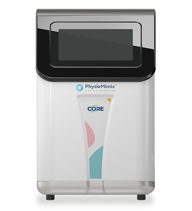
Add PhysioMimix Core in your lab
To develop your own cross-species Liver MPS models, you will need: