

Resource > Scientific publications >
Modeling Therapeutic Antibody-Small Molecule Drug-Drug Interactions Using a Three-Dimensional Perfusable Human Liver Coculture Platform
Filed under: ADME and Drug metabolism

Summary
The study used the LiverChip, CN Bio’s perfused three-dimensional (3D) liver microphysiological system (MPS) platform, to coculture primary human hepatocytes and Kupffer cells for 2 weeks and model a drug-drug interaction (DDI) between a therapeutic antibody and a small molecule under chronic interleukin-6 (IL-6)-mediated inflammation. IL-6 suppressed cytochrome P450 3A4 (CYP3A4) activity in a dose-dependent manner. The anti-IL-6 receptor antibody tocilizumab reversed that suppression, lowered C-reactive protein (CRP), and reduced exposure to the CYP3A4 substrate simvastatin hydroxy acid (SVA) by 19.0% to 21.0%, in the same direction as the interaction reported in patients with rheumatoid arthritis. Because conventional hepatocyte cultures lose metabolic function within days, the 2-week culture allowed measurement of both suppression and recovery of CYP3A4 activity in the same microtissues, which is relevant for evaluating therapeutic protein-drug interactions during drug development.
Study facts at a glance
| Publication | Long TJ, Cosgrove PA, Dunn RT II, Stolz DB, Hamadeh H, Afshari C, McBride H, Griffith LG. Modeling Therapeutic Antibody-Small Molecule Drug-Drug Interactions Using a Three-Dimensional Perfusable Human Liver Coculture Platform. Drug Metabolism and Disposition. 2016 Dec;44(12):1940-1948. |
| DOI | 10.1124/dmd.116.071456 |
| CN Bio product used | PhysioMimix® LC12 and PhysioMimix Core System |
| How the platform was used | Each bioreactor was seeded with 6.0 × 10⁵ cryopreserved primary human hepatocytes and 6.0 × 10⁴ Kupffer cells, and the resulting perfused 3D microtissues were cultured at a flow rate of 1 µL/s for 14 days, treated with interleukin-6 (IL-6) and tocilizumab across two sequential dosing periods, and dosed with simvastatin hydroxy acid (SVA) on days 7 and 12 for 48-hour pharmacokinetic (PK) sampling. |
| Biological context | Human hepatic drug metabolism under chronic IL-6-mediated inflammation, the inflammatory state associated with diseases such as rheumatoid arthritis. Cocultures of cryopreserved primary human hepatocytes and Kupffer cells (10:1) were run with three hepatocyte donor lots (HU8160, HU8163, and HU8196). Inflammation was induced with recombinant human IL-6 rather than with patient-derived disease cells; donor demographics are not specified in the main text. |
| Comparator | Within-platform controls (untreated cultures, IL-6 throughout, tocilizumab alone, and IL-6 washout) and before-versus-after tocilizumab measurements in the same cultures. Results were compared with published clinical tocilizumab-simvastatin data from patients with rheumatoid arthritis (Schmitt et al., 2011). No static or two-dimensional (2D) culture comparator was run in parallel. |
| Key readouts | Cytochrome P450 3A4 (CYP3A4) activity by luminescent assay; C-reactive protein (CRP), soluble IL-6 receptor (sIL-6R) alpha, albumin, and basal IL-6 by enzyme-linked immunosorbent assay (ELISA); SVA concentrations by liquid chromatography with tandem mass spectrometry (LC-MS/MS), with SVA half-life and area under the curve up to the last measurable concentration (AUClast); and confocal imaging of microtissue morphology. |
| Main interpretation | The perfused hepatocyte and Kupffer cell coculture remained stable for 2 weeks, long enough to capture IL-6-driven CYP3A4 suppression followed by tocilizumab-driven recovery in the same cultures, and it reproduced the direction of the clinically reported tocilizumab-simvastatin interaction, although the reduction in SVA exposure (19.0% to 21.0%) was smaller than the 39% reported clinically. |
Table of Contents
Which CN Bio product was used?
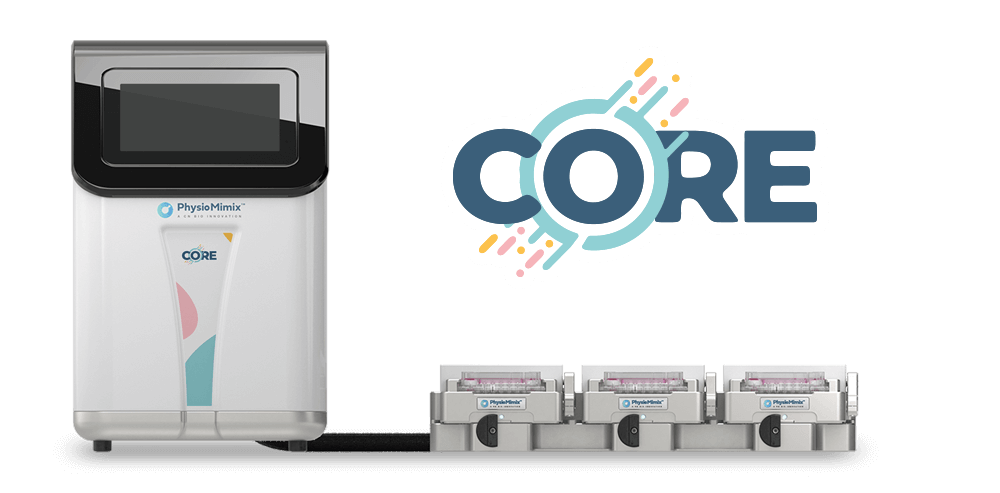
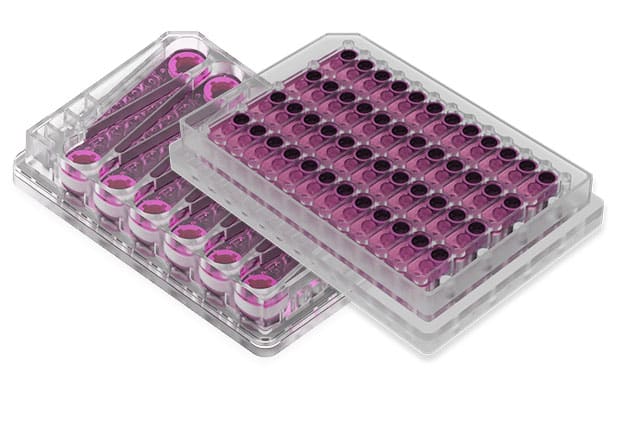
The study used the LiverChip, a CN Bio platform for perfused 3D liver culture and the forerunner of the PhysioMimix Core system. Each LiverChip platform holds 12 fluidically isolated bioreactors, formed by a polysulfone cell culture plate secured to a pneumatic plate. Each bioreactor contains a collagen-coated polystyrene scaffold, 1 cm in diameter, with 300 cylindrical microchannels (300 µm in diameter and 250 µm in height) where cells form 3D microtissues. Pneumatic pumping through a thin polyurethane membrane drives culture medium through the scaffold and recirculates it along a surface channel. In CN Bio’s current liver-on-a-chip format, primary human liver cells are cultured on the PhysioMimix Core system in the Multi-chip Liver-12 plate.
Each bioreactor was seeded with 6.0 × 10⁵ cryopreserved primary human hepatocytes and 6.0 × 10⁴ Kupffer cells in 1.6 mL of seeding medium, and cultures were kept at 37°C with 5% CO₂ at a flow rate of 1 µL/s. Flow ran downward for the first 8 hours to direct cells into the microchannels, then upward for the rest of the experiment to reoxygenate the medium along the surface channel. From culture day 3, cultures received Williams’ E maintenance medium containing 100 nM hydrocortisone, changed every 48 hours; dexamethasone was not used. All wells in the interaction study were induced with 10 µM rifampicin from day 5 to increase the dynamic range of CYP3A4 activity. IL-6 was used at 1 ng/mL and tocilizumab at 1.6 µM, a concentration chosen to represent the human serum maximum concentration (Cmax), with 72 hours of tocilizumab treatment before the second SVA dose. Cultures were maintained for 14 days.
The LiverChip supported all cell culture work, including IL-6 and tocilizumab treatments and SVA dosing. CYP3A4 activity, protein biomarkers, and SVA concentrations were measured offline from collected medium by luminescence, enzyme-linked immunosorbent assay (ELISA), and liquid chromatography with tandem mass spectrometry (LC-MS/MS), and scaffolds were removed at the end of culture for confocal imaging.
Find out more about CN Bio Liver-on-a-chip models here
Find out more about CN Bio DILI assays here
What this paper is about
Inflammation reduces the activity of several cytochrome P450 (CYP) enzymes in the liver, and CYP3A4 metabolizes over 50% of marketed small-molecule drugs. Chronic inflammatory diseases such as rheumatoid arthritis raise circulating IL-6, which suppresses hepatic CYP3A4. When a biologic such as tocilizumab, an anti-IL-6 receptor monoclonal antibody, blocks IL-6 signaling, CYP3A4 activity can recover and exposure to co-administered small molecules can fall. This has been observed clinically: tocilizumab reduced exposure to simvastatin (SVS) and its active form SVA in patients with rheumatoid arthritis (Schmitt et al., 2011). U.S. Food and Drug Administration (FDA) draft guidance from 2012 encouraged the exploration of these large-molecule/small-molecule DDIs to support regulatory filings and improve drug safety evaluation.
Studying this type of interaction in vitro requires hepatocytes that stay metabolically competent long enough to show two phases in sequence: suppression of CYP activity by a cytokine, then recovery when the cytokine signal is blocked. Primary human hepatocytes on flat, collagen-coated plates lose key functions, such as metabolic activity, after only a few days. Earlier two-dimensional (2D) work showed IL-6-mediated modulation of CYP activity. Still, it was limited to 96 hours of treatment or less, and it codosed an anti-IL-6 antibody with IL-6 to prevent suppression rather than reverse it (Dickmann et al., 2011). According to the authors, human hepatocyte data from the LiverChip had previously been limited to 1 week of culture.
This study, a collaboration between Amgen, the Massachusetts Institute of Technology (MIT), and the University of Pittsburgh, set out to reproduce the tocilizumab-SVA interaction in a perfused liver MPS. The researchers first characterized the dose response of CYP3A4, CRP, and soluble IL-6 receptor (sIL-6R) to IL-6, then ran a 14-day study with six treatment groups across two sequential dosing periods, measuring SVA pharmacokinetics (PK) before and after tocilizumab treatment in the same cultures, and repeated the design with three hepatocyte donors. Kupffer cells were included to build a more complete hepatic cellular environment, an approach also used in a 3D human liver-immunocompetent coculture study of diclofenac. CN Bio Innovations staff are acknowledged for LiverChip installation, hepatocyte lot qualification, and platform optimization, and the work was partly funded by the Defense Advanced Research Projects Agency (DARPA) and the National Institutes of Health (NIH). The application area is in vitro drug metabolism and inflammation-mediated DDIs.
What the researchers found
The study reported the following, in order of importance.
Tocilizumab reversed IL-6-driven suppression of CYP3A4 activity. CYP3A4 activity was expressed as the ratio between the end of the second dosing period (day 14) and the end of the first (day 9). In cultures treated with IL-6 and then with IL-6 plus tocilizumab, CYP3A4 activity increased 2.0 ± 0.5-fold for donor HU8160 and 1.7 ± 0.1-fold for donor HU8163 (both P < 0.05). Removing IL-6 after the first dosing period (washout) produced a similar recovery, with ratios of 1.9 ± 0.5 for HU8160 and 1.9 ± 0.1 for HU8163 (both P < 0.05). Activity ratios stayed close to 1 in untreated cultures, in cultures exposed to IL-6 throughout, in cultures that received IL-6 and tocilizumab together only in the second period, and in cultures that received tocilizumab alone. The authors interpret this pattern as showing that tocilizumab blocked IL-6 signaling in the culture and that its effect depended on the presence of an inflammatory cytokine.
Tocilizumab reduced SVA exposure in inflamed cultures. Compared with IL-6 treatment alone, adding tocilizumab after an initial IL-6 treatment reduced the SVA half-life ratio by 29.8% and the ratio of area under the curve up to the last measurable concentration (AUClast) by 21.0% for donor HU8160, and by 20.6% and 19.0% for donor HU8163 (all P < 0.05). In the clinical study, tocilizumab reduced SVA AUClast by 39% (90% confidence interval [CI], 24% to 51%) and SVS AUClast by 57%, with no significant change reported in SVA half-life (5 ± 1 hours before and 4 ± 2 hours after tocilizumab). The reduction in AUClast measured in the LiverChip was statistically significant compared with controls but fell just below the lower bound of the clinical 90% CI. The authors suggest the smaller effect may reflect the SVA concentration used (2.1 µM), which was well below its Michaelis constant (Km) of 50 to 80 µM and therefore implied a relatively low enzyme activity rate; they chose this concentration to balance physiologic relevance with the LC-MS/MS detection range.
IL-6 suppressed CYP3A4 activity and raised CRP in a dose-dependent manner. In cultures from donor HU8160, over 96 hours of IL-6 exposure (1 to 10,000 pg/mL) reduced CYP3A4 activity along a sigmoidal curve, with a half-maximal effective concentration (EC50) of 463 pg/mL. CRP secretion rose with an EC50 of 354 pg/mL, and sIL-6R levels decreased in a first-order manner with increasing IL-6, further evidence of IL-6 bioactivity in the cultures. Both EC50 values fell within the clinical range of IL-6 reported in inflammatory diseases (10 to 1500 pg/mL), and 1 ng/mL IL-6 was chosen for the chronic inflammation studies to saturate the response.
Inflammation lengthened SVA half-life and increased exposure. In untreated HU8160 cultures, SVA half-life was 5.0 ± 0.3 hours on day 7 and 4.9 ± 0.3 hours on day 12, and AUClast was 4841.0 ± 142.8 ng·h/mL and 4961.2 ± 127.5 ng·h/mL, respectively. Under 1 ng/mL IL-6, the mean SVA half-life increased to 6.4 ± 1.2 hours and 6.8 ± 1.5 hours in the two dosing periods, and AUClast increased to 5688.7 ± 831.2 ng·h/mL on day 7 and 6034.7 ± 889.7 ng·h/mL on day 12, consistent with reduced CYP3A4 activity.
CRP tracked the resolution of inflammation. In cultures from donor HU8160, IL-6 produced an initial 4-fold increase in CRP, which remained high in cultures dosed with IL-6 throughout. Tocilizumab reduced CRP within 2 days, reaching near-basal levels 3 days after antibody addition, and removing IL-6 produced the same pattern. Cultures with no inflammatory stimulus showed CRP levels that decreased toward 0 over time. The CRP results were corroborated with donor HU8163, where fold changes were larger because basal CRP was lower.
The coculture remained stable over 2 weeks. Albumin production was low before day 3, then increased and stabilized by day 5. Basal IL-6 started high (96.0 ± 46.5 pg/mL) and fell to healthy human serum levels (below 10 pg/mL) by day 7, which the authors interpret as an initial adaptation period after seeding. By day 7, microtissues several cell layers thick had formed inside the scaffold channels and extended through the 250 µm channel depth, with gaps that allowed fluid to flow through the scaffold.
One donor responded differently. In the third hepatocyte donor, HU8196, cultures treated with IL-6 alone unexpectedly showed CYP3A4 desuppression, giving an effect ratio close to that seen with tocilizumab. sIL-6R alpha levels in this donor approached 0 after 7 days of IL-6 treatment, which the authors suggest indicates that continued stimulation led to receptor loss and inconsistent IL-6 signaling, and tocilizumab treatment increased sIL-6R alpha levels in this donor. They conclude that for IL-6 to serve as a chronic inflammatory stimulus in long-term cultures, sIL-6R must not be depleted.
SVA handling and analytical constraints. SVA protein binding in the maintenance medium was 66%, and the 909 ng/mL SVA dose was set so that the unbound concentration was 10 times the reported human serum maximum concentration (Cmax) of 30.9 ng/mL for an 80 mg SVS dose. Interconversion of SVA to the simvastatin lactone form was negligible (below 1%). Researchers did not detect known phase I metabolites of SVA, likely because the large medium volume diluted their signals below background; the same medium volume also allowed repeated PK sampling over 48 hours.
Why the paper matters
For drug developers, the practical task with cytokine-modulating biologics is to anticipate how changes in inflammatory status will alter exposure to co-administered small molecules cleared by CYP3A4. These interactions are indirect, because tocilizumab changes CYP3A4 activity by interfering with the cytokine signaling that regulates CYP expression. Testing them in vitro therefore requires hepatocytes that respond to IL-6 and remain metabolically active for long enough to show suppression and recovery in sequence. This study shows that a perfused hepatocyte-Kupffer cell coculture in a liver MPS could sustain 2 weeks of culture and capture both phases, with SVA PK measured in the same cultures before and after tocilizumab. The authors describe the work as, to their knowledge, the first to directly demonstrate the clinically observed tocilizumab-SVA interaction in an in vitro liver culture model, made possible by the extended culture times of a 3D perfusion bioreactor.
The study also sets out a design that can be reused for other cytokine-mediated interactions: an IL-6 dose response to select a saturating inflammatory stimulus, two sequential dosing periods, and within-culture comparisons against untreated, inflamed, washout, and antibody-only controls. CN Bio’s immune-mediated liver injury models use the same principle, coculturing Kupffer cells with primary human hepatocytes to study inflammatory responses to small molecules and biologics. A cross-site reproducibility study of CN Bio’s liver MPS by U.S. FDA scientists also used lipopolysaccharide to challenge hepatocyte and Kupffer cell cocultures and reported CYP3A4 activity that remained stable for more than 2 weeks.
The authors place the work alongside computational approaches. Physiologically based pharmacokinetic (PBPK) models have been used to simulate IL-6-mediated CYP effects. Still, those models often rely on parameters generated from in vitro datasets, and the authors note that advanced in vitro systems combined with in silico modeling continue to improve early drug development. CN Bio discusses this combination further in its article on integrating in silico tools with organ-on-a-chip for absorption, distribution, metabolism, and excretion (ADME) studies.
The authors present the study as a proof of concept, and its scope is narrow. It tested one antibody, one small-molecule substrate, and one cytokine, with three technical replicates per group and three hepatocyte donors, one of which diverged because of sIL-6R depletion. The reduction in SVA exposure was smaller than the clinical effect; SVA metabolites could not be measured; and all cultures received rifampicin and hydrocortisone to maintain CYP3A4 activity (without hydrocortisone, basal CYP3A4 activity fell to low levels after 1 week). The authors identify other IL-6-mediated mechanisms, such as effects on drug transporter activity, as logical extensions and expect to apply these models beyond PK and toxicity evaluation to pharmacodynamic and human disease models.
Key study takeaways
- The study used CN Bio’s LiverChip platform to coculture primary human hepatocytes and Kupffer cells as perfused 3D microtissues for 14 days, modeling a therapeutic antibody-small molecule DDI under chronic IL-6-mediated inflammation.
- IL-6 suppressed CYP3A4 activity (EC50 of 463 pg/mL) and increased CRP secretion (EC50 of 354 pg/mL), with both EC50 values within the clinical range of IL-6 reported in inflammatory diseases.
- Tocilizumab added to IL-6-inflamed cultures increased CYP3A4 activity 1.7-fold to 2.0-fold, returned CRP to near-basal levels within 3 days, and reduced SVA AUClast by 19.0% to 21.0% compared with IL-6 alone in two hepatocyte donors.
- Compared with the published clinical study, the LiverChip reproduced the direction of the tocilizumab-SVA interaction. Still, the reduction in SVA exposure fell just below the lower bound of the clinical 90% CI (24% to 51%).
- The workflow combined CYP3A4 activity, CRP, sIL-6R, albumin, and IL-6 measurements with LC-MS/MS-based SVA PK sampled repeatedly over 48 hours in two dosing periods.
- The authors present the work as a proof of concept for modeling cytokine-mediated DDIs; donor-dependent receptor depletion, a smaller effect size than seen clinically, and a single antibody-substrate pair limit how far the results can be generalized.
Why this paper is worth reading
This paper is useful because it shows drug metabolism and pharmacokinetics (DMPK) and safety scientists how to design an in vitro study of a cytokine-mediated DDI in which suppression and recovery of CYP3A4 activity occur in the same cultures, with small-molecule PK measured before and after adding the biologic. It gives practical detail that can be reused directly: IL-6 EC50 values for CYP3A4 and CRP, the choice of a saturating 1 ng/mL IL-6 stimulus, the use of rifampicin and hydrocortisone to preserve CYP3A4 dynamic range, SVA dosing corrected for medium protein binding, and sIL-6R depletion as a donor-dependent pitfall. The comparison with clinical data also sets realistic expectations for how closely the magnitude of an in vitro interaction may match patient data. Teams planning inflammation-mediated DDI or biologic safety studies in a perfused liver model can run them in their own laboratory or through CN Bio’s absorption, distribution, metabolism, and excretion (ADME) study services, and CN Bio’s webinar on building regulatory confidence with immune-competent models covers how immune-competent liver MPS are being developed for monoclonal antibody safety assessment.
FAQ
The study used the PhysioMimix LC12 plate and the PhysioMimix Core System, both from CN Bio.
The LiverChip was used to coculture 6.0 × 10⁵ primary human hepatocytes and 6.0 × 10⁴ Kupffer cells per bioreactor as perfused 3D microtissues at 1 µL/s for 14 days. The cultures were treated with interleukin-6 (IL-6) and the anti-IL-6 receptor antibody tocilizumab in two sequential dosing periods and dosed with simvastatin hydroxy acid (SVA) on days 7 and 12 to measure SVA pharmacokinetics.
The study modeled human hepatic drug metabolism under chronic IL-6-mediated inflammation, the inflammatory state associated with rheumatoid arthritis, using cocultures of cryopreserved primary human hepatocytes and Kupffer cells from three hepatocyte donors. Inflammation was induced with recombinant human IL-6.
In the LiverChip coculture, tocilizumab reversed IL-6-driven suppression of cytochrome P450 3A4 (CYP3A4) activity. It reduced SVA exposure (AUClast) by 19.0% to 21.0% compared with IL-6 alone, in the same direction as the clinically reported tocilizumab-simvastatin interaction. The reduction was smaller than the 39% reported in patients with rheumatoid arthritis.
The study compared tocilizumab-treated LiverChip cultures with untreated, IL-6-only, tocilizumab-only, and IL-6 washout controls, and compared SVA pharmacokinetics in the same cultures before and after tocilizumab. The results were benchmarked against published clinical data from patients with rheumatoid arthritis; no static or 2D culture comparator was run in parallel.
The readouts were CYP3A4 activity, CRP, soluble IL-6 receptor, albumin, and basal IL-6 levels, SVA concentrations by LC-MS/MS with SVA half-life and AUClast, and confocal imaging of liver microtissue morphology.
The paper gives DMPK and drug safety teams a worked study design for testing cytokine-mediated drug-drug interactions in a perfused liver MPS, with suppression and recovery of CYP3A4 activity measured in the same cultures. It also shows the approach’s practical limits, including donor-dependent IL-6 receptor depletion and a smaller effect size than seen clinically.
Full citation
Long TJ, Cosgrove PA, Dunn RT II, Stolz DB, Hamadeh H, Afshari C, McBride H, Griffith LG. Modeling Therapeutic Antibody-Small Molecule Drug-Drug Interactions Using a Three-Dimensional Perfusable Human Liver Coculture Platform. Drug Metabolism and Disposition. 2016 Dec;44(12):1940-1948. DOI: 10.1124/dmd.116.071456.
Related products and services
Contract research services
Discover how to utilize our cross-species models to inform next-step decision making via our DILI in vitro Contract Research Services here.
Add PhysioMimix Core in your lab
To develop your own cross-species Liver MPS models, you will need: