

Resource > Scientific publications >
“Liver-on-a-Chip” Cultures of Primary Hepatocytes and Kupffer Cells for Hepatitis B Virus Infection
Filed under: Disease modeling and HBV
Summary
Researchers at Imperial College London used the CN Bio LiverChip perfused liver platform, the system now supplied as the PhysioMimix® Core with multi-chip liver plates, to establish 3D cultures of primary human hepatocytes (PHH), either as monocultures or in coculture with primary human Kupffer cells, and to infect them with purified hepatitis B virus (HBV). The published protocol covers plate assembly and equilibration, seeding of 600,000 PHH per well onto collagen-coated scaffolds under continuous perfusion at 1 µL/s, production and sucrose cushion purification of infectious HBV from inducible producer cell lines, infection at low multiplicity of infection (MOI), and the downstream analysis of viral and host responses. The representative results show hepatic microtissue formation within 3 days of seeding, stable albumin secretion to day 40 post-seeding, and HBV DNA detectable from day 2 post-infection at inocula as low as 0.05 genome equivalents (GE) per cell, without the dimethyl sulfoxide (DMSO) and polyethylene glycol (PEG) that conventional static cultures require. For groups working on hepatitis B, the paper provides a documented human in vitro route to long-term infection studies covering viral persistence, innate immune response, and sequential antiviral treatment.
Study facts at a glance
| Publication | Ortega-Prieto AM, Skelton JK, Cherry C, Briones-Orta MA, Hateley CA, Dorner M. “Liver-on-a-Chip” Cultures of Primary Hepatocytes and Kupffer Cells for Hepatitis B Virus Infection. Journal of Visualized Experiments. February 19, 2019;(144):e58333. |
| DOI | 10.3791/58333 |
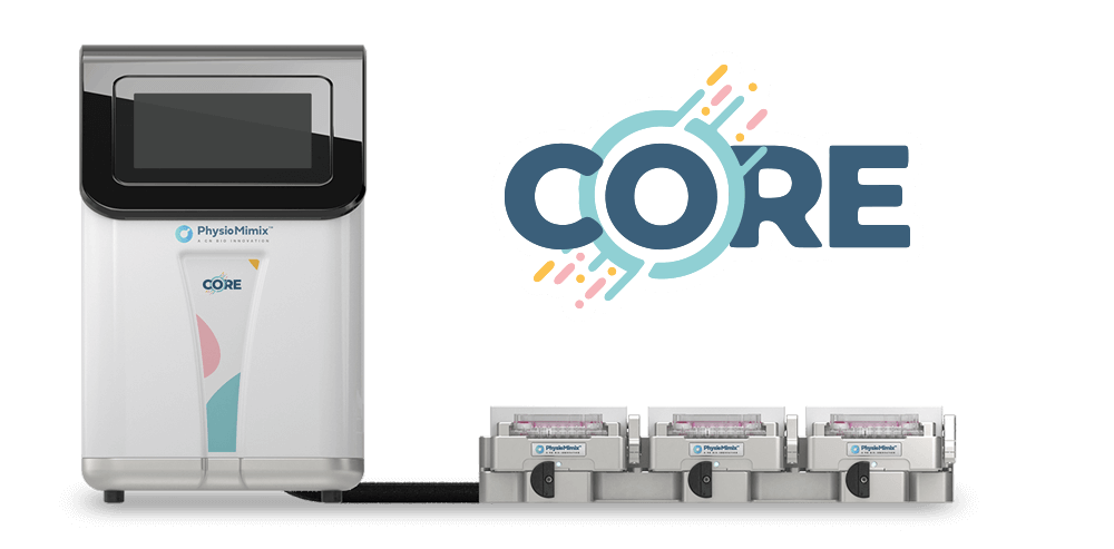
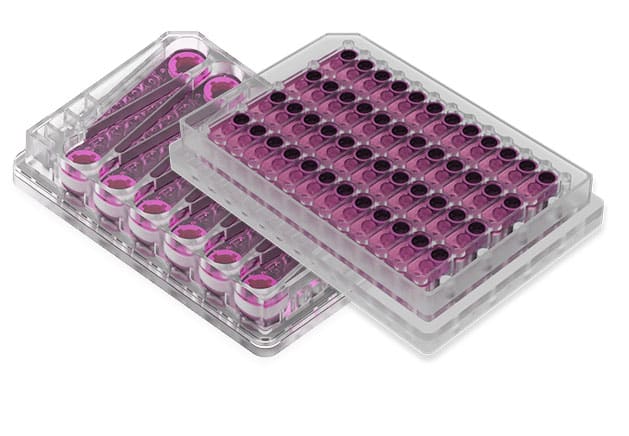
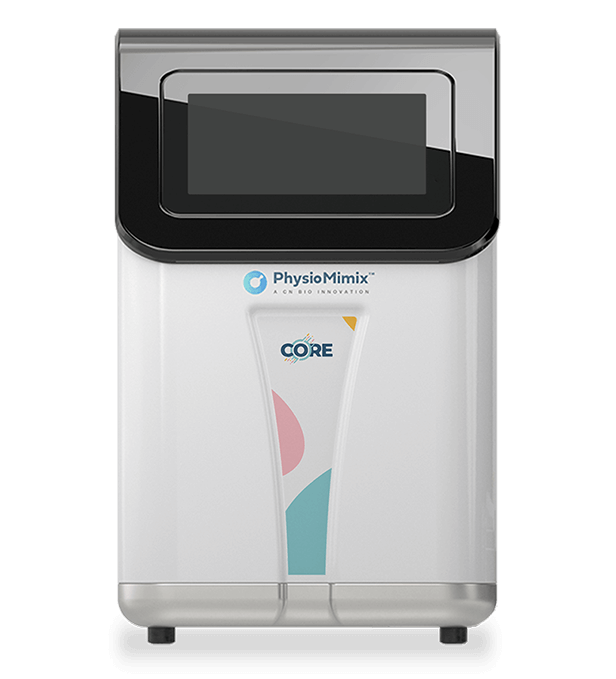
| CN Bio product used | PhysioMimix® Multi-chip Liver-12 plate and PhysioMimix Core System |
| How the platform was used | PHH were seeded at 600,000 cells per well onto collagen-coated scaffolds, alone or with 60,000 primary human Kupffer cells, then continuously perfused at 1 µL/s (8 h of downward flow followed by 8 h of upward flow during seeding) in a total well volume of 1.6 mL, comprising 1.4 mL in the well and 0.2 mL in the flow channel. Cultures were infected with purified HBV 3 days after seeding, with medium replaced every 48 h, and maintained for at least 40 days. |
| Biological context | Hepatitis B virus infection of human liver tissue. Cryopreserved primary human hepatocytes were cultured as 3D perfused microtissues, either alone or in coculture with primary human Kupffer cells, in a microphysiological system (MPS). Disease context was introduced by infection rather than being donor-derived. |
| Comparator | PHH monocultures were the specified control for PHH/Kupffer cell cocultures, mock-infected cultures were the control for infection, and conventional static 2D PHH cultures on collagen-coated plates served as the literature benchmark. Inocula spanning 0.05 to 5,000 GE per cell were tested. |
| Key readouts | Extracellular HBV DNA by quantitative polymerase chain reaction (qPCR), intracellular HBV pregenomic RNA (pgRNA) relative to the housekeeping gene RPS11, immunofluorescence detection of hepatitis B core antigen (HBcAg) and hepatitis B surface antigen (HBsAg), human albumin by enzyme-linked immunosorbent assay (ELISA), interleukin 6 (IL6) and tumor necrosis factor alpha (TNFα) following lipopolysaccharide (LPS) stimulation, and brightfield imaging of microtissue formation. |
| Main interpretation | The protocol sets out a reproducible route to long-term perfused PHH and PHH/Kupffer cell cultures that support HBV infection at low multiplicity of infection without DMSO or PEG, giving access to viral, hepatic function, and innate immune readouts across culture periods of at least 40 days. |
Table of Contents
Which CN Bio product was used?
The study used the perfused liver platform that the authors call the LiverChip, operated with its compressor, vacuum pump, washing dock, and docking station. Microfluidic plates were assembled by seating a sterile membrane on the base plate, adding the well-containing top plate and lid, and tightening the screws symmetrically to 33 lb by automated precision torque and 35 lb by manual torque. Each plate was primed with 400 µL of hepatocyte seeding medium per well and run at 1 µL/s in the upward direction for 3.5 min, with the red indicators at the side of the plate confirming that microfluidic circulation had been established. A further 1.2 mL of seeding medium was added and the plate was equilibrated overnight at 1 µL/s upward flow for 16 h at 37 °C and 5% CO2 before a filter paper, a cell attachment scaffold, and a retaining ring were locked into each well.
Cells were then seeded directly onto the collagen-coated scaffolds, which are continuously perfused with growth medium to supply oxygen and nutrients. Monocultures received 600,000 PHH per well in 500 µL of hepatocyte seeding medium (Williams E medium with primary hepatocyte thawing and plating supplements, 5% fetal bovine serum, and 1 µM dexamethasone). Cocultures received 60,000 Kupffer cells and 600,000 hepatocytes in 250 µL each of coculture seeding medium, prepared in advanced Dulbecco’s modified Eagle’s medium without dexamethasone. Seeding in both formats used 8 h of downward flow at 1 µL/s followed by flow reversal to the upward direction for 8 h, and maintenance medium was replaced every 48 h with a washing step to ensure complete medium exchange. The authors note that the plate format holds 12 scaffolds, that this can be upgraded to 36, and that scaling to higher well numbers was not possible at the time of publication because of the microfluidic requirements of the platform.
The organ-on-a-chip platform covers the culture and infection phases of the workflow. Production of infectious HBV from inducible producer cell lines, PEG precipitation and sucrose cushion purification, qPCR quantification of HBV DNA and pgRNA, albumin ELISA, immunofluorescence staining, and cytokine measurement were all performed off-platform using standard laboratory equipment, with virus production and infection steps carried out in a containment level III laboratory.
Find out more about CN Bio Liver-on-a-chip models here
Find out more about CN Bio DILI assays here
What this paper is about
HBV research has long been constrained by the poor susceptibility of culture systems. The authors set the problem out in terms of infectivity: as few as three viral genomes can result in chronicity in experimentally infected chimpanzees in vivo, whereas most in vitro models require several hundred to several thousand viral genomes per cell to initiate an infection that is only transient. The second constraint is longevity. Primary human hepatocytes are fragile and dedifferentiate rapidly in conventional culture, which the authors attribute to flat, hard plastic surfaces that do not reproduce the extracellular environment of the liver and to the general lack of oxygenation in the absence of microfluidic circulation. Conventional static hepatocyte cultures on collagen-coated plates lose their susceptibility to HBV infection as they dedifferentiate.
The protocol addresses both constraints with a 3D liver-on-a-chip model in which PHH are seeded onto collagen-coated scaffolds and continuously perfused, supporting cultures of at least 40 days. Two configurations are described in parallel: PHH monocultures, and cocultures of PHH with primary human Kupffer cells, the resident macrophage population of the liver. The paper also describes the production of purified HBV suitable for low-multiplicity-of-infection studies, and the analysis of both viral and host responses.
The comparison the authors draw is with other in vitro systems. They note that alternative PHH culture systems based on complex cocultures of murine fibroblasts, or on 3D growth in spheroids, have been validated and are susceptible to HBV infection at multiplicities of 500 GE per cell, and that establishing infection in these cultures depends on high concentrations of DMSO and PEG. Their position, referenced to their earlier work, is that 3D liver-on-a-chip cultures were at the time the only in vitro system reported to be susceptible at 0.05 GE per cell, and that DMSO and PEG are dispensable in this format. The applications named for the platform are drug efficacy studies, toxicological analysis, investigation of pathogenesis, evaluation of curative therapies aimed at eliminating covalently closed circular DNA (cccDNA), and assessment of sequential drug treatments and their effect on HBV persistence.
This is a protocol paper, and the representative results shown are adapted from the same group’s earlier study in Nature Communications (Ortega-Prieto AM et al., 2018;9:682). The work was funded by a European Research Council Starter grant, a Wellcome Trust Investigator Award, and CN Bio Innovations.
What the researchers found
The representative results show that hepatic microtissues form within 3 days of seeding PHH, with functional bile canaliculi and complete cell polarization. Human albumin, which the authors describe as the best marker for evaluating hepatic metabolism, was stably and highly expressed by the 3D cultures until day 40 post-seeding, measured as both total and per-cell adjusted production by ELISA.
The infection data show that these cultures become highly susceptible to HBV. HBV DNA and other viral markers were readily detectable in culture supernatants from day 2 post-infection, in contrast to other culture systems. The paper reports susceptibility across a range of inocula, and states that where conventional hepatocyte cultures require at least 500 HBV GE per cell together with 2% DMSO and 4% PEG, as few as 0.05 GE per cell were sufficient to initiate infection in the 3D cultures with neither additive. Intracellular pgRNA accumulated relative to RPS11 between day 2 and day 10, and immunofluorescence staining of scaffolds retrieved from the cultures detected HBcAg and HBsAg 10 days after infection.
Kupffer cell function was assessed by cytokine secretion. Cocultures were treated with 1 µg/mL LPS at day 9 post-seeding for 48 h and the medium was harvested at day 11. IL6 secretion rose sharply in LPS-stimulated cocultures relative to unstimulated cocultures and to hepatocyte monocultures, and TNFα was detected only in the cocultures. The authors recommend capture-based detection for these measurements and advise lot testing of Kupffer cells, since commercially available donors differ in their responsiveness to LPS stimulation. This gives the coculture format a functional readout for the innate immune compartment alongside the hepatocyte and viral endpoints.
The discussion adds several operational findings that determine whether the protocol works. The most critical step named is the quality of the sourced primary cells: only plateable PHH lots that have been tested for adherence capacity should be used, and although freshly isolated PHH can be used, their cryopreservation is complicated and requires rate-controlled freezers. Viral inoculum preparation matters just as much. PEG-precipitated and sucrose cushion-purified HBV is required when using inducible producer cell lines, because cell culture supernatants applied directly to the cultures do not readily result in infection, either through inhibitory factors or through incompatibility of the growth factors present with hepatocytes. Where patient-derived inocula are used, only serum is suitable, since plasma coagulates and clogs the microfluidic circulation.
Two further points affect experimental design. Dosing calculations must account for the microfluidic channel as well as the well, so the 1.4 mL well volume and the 0.2 mL channel volume are both included, and one washing step with drug-containing or virus-containing medium is performed to prime the channel. The authors also report that, in contrast to conventional static 2D cultures, host genetic background was negligible with regard to HBV susceptibility, with all hepatocyte donors tested to date able to establish infection. They are explicit about one limitation shared with other platforms: once established, HBV infection does not readily spread to uninfected cells, and the mechanism for this remains unresolved given that HBV infects the majority of hepatocytes in vivo.
Why the paper matters
For virology and antiviral groups, the practical value sits in the combination of inoculum economy and culture duration. Infecting at 0.05 GE per cell without DMSO or PEG removes the need for large volumes of purified virus and for additives that other systems depend on, and a culture window of at least 40 days makes it feasible to run sequential drug treatments and to follow HBV persistence over a timescale that conventional hepatocyte cultures cannot support. Studies aimed at cccDNA, the transcriptional template for all newly produced virions, need exactly that kind of window.
The Kupffer cell coculture is the second point. Adding a resident macrophage population to a perfused hepatocyte culture gives access to cell-to-cell interactions and to host response biomarkers in a system where hepatocytes remain metabolically competent, which supports disease modeling work on pathogenesis as well as on antiviral efficacy. The paper also documents the failure modes that decide whether the experiment succeeds, including cell lot quality, inoculum purification, serum rather than plasma for patient-derived virus, and volume accounting for dosing, so these do not have to be rediscovered.
There is a translational argument here too. HBV is human-tropic, which places a practical limit on how far the questions in this paper can be answered in other species, and the protocol provides a human in vitro route to studying infection, host response, and treatment in liver tissue that retains metabolic and functional competence. All models have strengths and limitations, and the authors are clear about where this one stops, but for long-term HBV infection studies the protocol offers a documented alternative to shorter-lived static systems.
Key study takeaways
- The study used the CN Bio perfused liver platform, referred to in the paper as the LiverChip, to culture 600,000 PHH per well on collagen-coated scaffolds under continuous perfusion at 1 µL/s, alone or with 60,000 primary human Kupffer cells.
- The cultures formed hepatic microtissues within 3 days of seeding, with functional bile canaliculi and complete cell polarization, and maintained stable albumin secretion to day 40 post-seeding.
- Compared with conventional hepatocyte cultures, which the authors state require at least 500 HBV GE per cell plus 2% DMSO and 4% PEG, the 3D cultures were infected with as few as 0.05 GE per cell without either additive.
- The workflow combined extracellular HBV DNA qPCR, intracellular pgRNA relative to RPS11, immunofluorescence detection of HBcAg and HBsAg, albumin ELISA, and IL6 and TNFα measurement after LPS stimulation.
- LPS-stimulated PHH/Kupffer cell cocultures secreted IL6 and TNFα where hepatocyte monocultures did not, giving the coculture format a functional readout for Kupffer cell viability and responsiveness.
- The paper indicates the model is most appropriate for long-term HBV infection studies, treatment combinations, and pathogenesis, and notes that established infection does not readily spread to uninfected cells and that the plate format holds 12 scaffolds, upgradeable to 36.
Why this paper is worth reading
This paper is useful because it publishes the operational detail behind a long-term HBV infection experiment rather than only its conclusions: torque settings, priming and equilibration times, medium compositions for monoculture and for both stages of coculture, flow directions and durations at each step, cell numbers per well, and the qPCR, ELISA, and staining conditions used for each readout. As with the later MPS protocol for modeling fractionated radiotherapy in rectal tumor organoid-derived monolayers, the value lies in parameters a group can plan against before committing donor material. A scientist deciding whether to build an HBV infection model in a perfused liver MPS can use it to judge how much purified virus a study will consume, how long the cultures will hold, which Kupffer cell and hepatocyte lots to qualify in advance, and how to account for the microfluidic channel volume when dosing. The troubleshooting content is worth attention on its own, since the guidance on inoculum purification, serum rather than plasma, and plateable cell lots addresses the failure modes most likely to cost a study its first run. Groups without in-house MPS capacity can access equivalent liver models through CN Bio contract research services.
FAQ
The study used the PhysioMimix® Core system with the Multi-chip Liver-12 plate. The plate provides 12 perfused, scaffold-based liver chips.
Primary human hepatocytes were seeded at 600,000 cells per well onto collagen-coated scaffolds in the microfluidic plate, alone or with 60,000 primary human Kupffer cells, and continuously perfused at 1 µL/s in a total well volume of 1.6 mL. Cultures were infected with purified HBV 3 days after seeding and maintained for at least 40 days, with medium exchanged every 48 h.
The disease area is hepatitis B virus infection of the human liver. The model comprises cryopreserved primary human hepatocytes cultured as 3D perfused hepatic microtissues, either as monocultures or in coculture with primary human Kupffer cells.
The protocol establishes long-term perfused cultures of primary human hepatocytes and Kupffer cells that support HBV infection at as few as 0.05 genome equivalents per cell without DMSO or PEG, with HBV DNA detectable from day 2 post-infection. The cultures formed hepatic microtissues within 3 days and maintained stable albumin secretion to day 40 post-seeding.
PHH monocultures were compared with PHH/Kupffer cell cocultures, infected cultures were compared with mock-infected controls, and inocula spanning 0.05 to 5,000 genome equivalents per cell were compared for susceptibility. Conventional static 2D primary human hepatocyte cultures on collagen-coated plates, along with micropatterned cocultures and spheroids, were used as the literature benchmark.
The readouts were extracellular HBV DNA by qPCR, intracellular HBV pgRNA relative to RPS11, immunofluorescence detection of HBcAg and HBsAg, human albumin by ELISA, IL6 and TNFα after LPS stimulation, and brightfield imaging of hepatic microtissue formation.
The paper gives researchers a documented protocol for long-term HBV infection studies in a perfused human liver MPS, including the cell numbers, flow rates, medium compositions, infection conditions, and analytical methods needed to plan an experiment. It also states where the model stops being appropriate, noting that established HBV infection does not readily spread to uninfected cells and that the platform cannot currently be scaled beyond 36 scaffolds per plate.
Full citation
Ortega-Prieto AM, Skelton JK, Cherry C, Briones-Orta MA, Hateley CA, Dorner M. “Liver-on-a-Chip” Cultures of Primary Hepatocytes and Kupffer Cells for Hepatitis B Virus Infection. Journal of Visualized Experiments. February 19, 2019;(144):e58333. DOI: 10.3791/58333.
Related products and services
Contract research services
Discover how to utilize our cross-species models to inform next-step decision making via our DILI in vitro Contract Research Services here.
Add PhysioMimix Core in your lab
To develop your own cross-species Liver MPS models, you will need: