

Resource > Scientific publications >
Dynamic Culture Improves the Predictive Power of Bronchial and Alveolar Airway Models of SARS-CoV-2 Infection
Filed under: Disease modeling and Pulmonary infection
Summary
Researchers at the Liverpool School of Tropical Medicine, working with CN Bio, used the PhysioMimix Core system to build perfused bronchial and alveolar lung-on-a-chip models from primary human airway epithelial and pulmonary endothelial cells, then compared them directly with matched static Transwell cultures during infection with three SARS-CoV-2 variants. Dynamic flow produced pseudostratified bronchial epithelium and alveolar sac-like structures with higher expression of region-specific cell markers, and yielded more differentially expressed genes after infection because fold changes were larger and variability between replicates was lower. For groups selecting a model for respiratory infection work, the study indicates that static bronchial cultures can suffice for basic infection dynamics. In contrast, perfused models add resolution when transcriptomic endpoints or alveolar infection are the objective.
Study facts at a glance
| Publication | Caygill CH, Lopeman R, Lewis KA, Richardson E, Casas Sanchez A, Heavey N, Winrow A, Howard L, Williams C, Wooding D, Edwards T, Lucas E, Kostrzewski T, Owen A, Pennington SH, Biagini GA. Dynamic Culture Improves the Predictive Power of Bronchial and Alveolar Airway Models of SARS-CoV-2 Infection. bioRxiv. 2025 Jul 21. Preprint, not certified by peer review. |
| DOI | 10.1101/2025.07.21.665885 |
| CN Bio product used | PhysioMimix® Multi-chip Barrier-12 plate and PhysioMimix Core System |
| How the platform was used | Primary human bronchial or small airway epithelial cells were seeded on the apical surface of collagen I-coated Transwell® inserts, with human pulmonary microvascular endothelial cells on the basolateral surface. Inserts were placed in Multi-chip Barrier-12 plates in the PhysioMimix docking station, perfused at 0.5 µL/s (to approximate human blood flow), and differentiated at the air-liquid interface (ALI) for 14 to 16 days before challenge or infection. |
| Biological context | The human respiratory tract was modeled in two anatomically distinct regions. Bronchial models used normal human bronchial epithelial cells (NHBE); alveolar models used small airway epithelial cells (SAEC). Both were co-cultured with human pulmonary microvascular endothelial cells (HPMEC). All cells were commercially sourced primary cells from healthy donor material. Disease context was acute SARS-CoV-2 infection (COVID-19). |
| Comparator | Matched static Transwell cultures: the same primary cells, inserts, media, and differentiation protocol, held in standard 24-well plates without perfusion. Secondary comparisons covered epithelial monoculture versus epithelial-endothelial co-culture, bronchial versus alveolar models, and three SARS-CoV-2 variants (Pre-alpha, Delta B.1.617.2, Omicron BA.5). |
| Key readouts | Histology (hematoxylin and eosin, Alcian blue), transepithelial electrical resistance (TEER), qPCR for lineage markers and for the entry receptors ACE2 and TMPRSS2, immunofluorescence for cilia, mucus, surfactant, and SARS-CoV-2 nucleocapsid protein, IP-10 secretion by ELISA following poly(I:C) and lipopolysaccharide (LPS) challenge, infection rate, viral RNA by qPCR, infectious titer by Vero E6 plaque assay, and NanoString gene expression (785 host response targets plus 48 custom lung targets) with pathway global significance scoring. |
| Main interpretation | Perfusion improved airway tissue architecture, regional marker expression, and the sensitivity and reproducibility of transcriptomic readouts, while infection rates in bronchial tissues were comparable between static and perfused culture; therefore, the reported benefit of dynamic flow rests on data quality and alveolar permissiveness rather than infection efficiency alone. |
Table of Contents
Which CN Bio product was used?
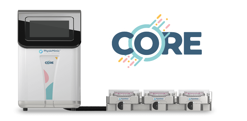
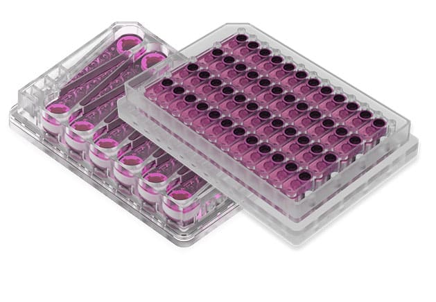
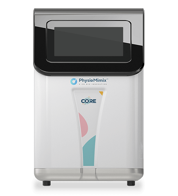
The study used the PhysioMimix Core system, comprising the docking station and driver, together with Multi-chip Barrier-12 plates (named MPS-T12 plates in the publication). Primary human bronchial epithelial cells or small airway epithelial cells were seeded apically onto collagen I-coated 24-well, 6 mm, 0.4 µm pore PET Transwell inserts, with pulmonary microvascular endothelial cells seeded on the inverted basolateral surface. Flow was introduced 24 hours after seeding at 0.5 µL/s and maintained through a 14-16-day differentiation period at ALI, with basolateral media exchanged every 48 to 72 hours and apical PBS washes to clear accumulated mucus.
The platform was used to generate and mature the tissues rather than to house every experiment. The publication states that plates were removed from the docking station at the end of differentiation and transferred, alongside the static plates, to an incubator in a containment level 3 laboratory for SARS-CoV-2 infection and the subsequent seven-day sampling period. The infection comparisons therefore contrast tissues conditioned by perfusion during differentiation against tissues never exposed to it. Static comparator cultures were held in standard 24-well plates.
Find out more about CN Bio Lung-on-a-chip models here
What this paper is about
Microphysiological systems (MPS) are widely proposed as human-relevant alternatives to conventional preclinical models. Still, the authors note that comparative data benchmarking perfused models against their static counterparts remains scarce, and that a published meta-analysis found only modest improvements in biomarker expression in perfused tissue. In infectious disease research specifically, several groups have shown that MPS support replication of respiratory pathogens. Yet, the authors state that none had formally tested whether the added complexity delivers improved functional performance relative to static systems.
This study addresses that question directly. Bronchial and alveolar airway models were built on the same insert format, with the same primary cells and the same differentiation protocol, and split between perfused and static conditions so that flow was the only variable. The models were then characterized for tissue morphology, lineage marker expression, barrier integrity, and innate immune responsiveness, and infected with three SARS-CoV-2 variants spanning the early pandemic and later immune-evasive lineages. SARS-CoV-2 was chosen as the model pathogen because its tropism and clinical outcomes differ across the respiratory tract, allowing region-specific model performance to be assessed against known clinical observations. Readers can find the underlying culture format described in CN Bio’s lung-on-a-chip technology explainer.
What the researchers found
The study reported that dynamic flow improved tissue architecture in both airway regions. Bronchial cultures under perfusion formed a pseudostratified epithelium resembling native bronchial tissue, while static cultures appeared less developed. Alveolar cultures under perfusion formed protruding sac-like structures with air pockets, whereas static alveolar cultures remained flattened, uniform cell layers. Marker expression followed the same pattern: club cell (SCGB1A1) and goblet cell (MUC5AC) transcripts were significantly higher in perfused bronchial tissues, and alveolar type I (AQP5) and type II (SFTPB) markers were better maintained in perfused alveolar tissues over 14 days.
Barrier measurements did not separate the two conditions. TEER showed no significant difference between static and perfused bronchial tissues, and no significant difference between conditions in either co-culture model. Adding endothelial cells increased TEER in both models, with peak values of 1859.82 Ω·cm² in bronchial and 1766.23 Ω·cm² in alveolar co-cultures, and supported retention of differentiated epithelial phenotypes under flow.
Transcriptomic comparison found no significant gene expression differences between static and perfused tissues within the same airway region. The difference emerged between regions: 23 lung region-specific genes were differentially expressed between bronchial and alveolar models, and bronchial markers including MUC5B, BPIFB1, MUC5AC, and SCGB1A1 separated the two regions significantly under dynamic flow but not under static culture. The authors attribute the larger number of differentially expressed genes under flow primarily to the magnitude of fold changes.
Perfused co-cultures responded to innate immune stimuli in a tissue-appropriate way. Both bronchial and alveolar models secreted IP-10 within six hours of apical poly(I:C) challenge and sustained the response over 48 hours, with alveolar tissues releasing more IP-10 than bronchial tissues. LPS increased IP-10 only in the alveolar model, which the authors relate to low TLR4 expression in bronchial epithelium. ACE2 expression was significantly higher in bronchial than alveolar tissues, while TMPRSS2 was comparable, matching the clinical observation that ACE2 expression decreases down the respiratory tract. Permissiveness was confirmed with a SARS-CoV-2 D614G pseudotyped lentivirus.
For infection itself, results depended on region and variant more than on flow. At a multiplicity of infection (MOI) of 1, bronchial infection rates were high in both conditions, with no significant difference between static and perfused conditions for any variant. Alveolar tissues were more variable: Omicron BA.5 reached 100% infection in both systems, while Pre-alpha infected 47% of perfused and 43% of static alveolar tissues, and Delta infected 69% of perfused and 43% of static alveolar tissues. At an MOI of 0.01, Pre-alpha infected 44% of perfused bronchial tissues and 0% of static bronchial tissues (p = 0.022), and alveolar tissues did not support infection in either system. Viral titers peaked at day four across all variants. Pre-alpha replicated 10- to 100-fold higher in bronchial than alveolar tissues, Delta 6- to 10-fold higher, and Omicron BA.5 replicated to similar levels in both regions. Immunofluorescence showed Delta producing widespread infection with large syncytia and punctate nuclear staining indicative of DNA damage, while Omicron BA.5 infection localized to discrete concentrated foci.
Host response analysis placed interferon signaling at the center of the response. Type I, II, and III interferon pathways were the most significantly enriched across all models and variants. Delta infection produced the most differentially expressed genes and the largest overall fold changes, along with pathway activations not seen in other variants, including proteotoxic stress, oxidative stress, leukotriene and prostaglandin signaling, and IL-2 signaling. Perfused bronchial cultures returned more differentially expressed genes than static bronchial cultures, and plotting log₂ fold change against coefficient of variation showed this was driven by both larger effect sizes and reduced variability across replicates. Global significance scores could not be generated for static bronchial tissues infected with Pre-alpha or Omicron BA.5 because too few genes met the significance threshold.
Why the paper matters
The practical value is model-selection guidance grounded in a controlled comparison, not a general claim about perfusion. The authors are explicit that for studies assessing infection dynamics alone, static bronchial models may be sufficient, and they scope that conclusion to SARS-CoV-2 while noting other pathogens may behave differently. Perfused models earn their place in discrete quantitative analyses: greater fold changes and lower replicate variability mean more differentially expressed genes reach significance, which the authors link to greater confidence from the same experiment and fewer replicates required. That argument carries weight in work built on primary and donor cells, which are costly and available in limited quantity.
The regional separation matters for study design. Because bronchial and alveolar models differ in ACE2 expression, cytokine output, and permissiveness to individual variants, matching the model to the research question becomes a consequential decision: bronchial tissues suit work on primary infection sites. In contrast, alveolar tissues suit disease progression and pathogenicity, as they are associated with acute respiratory distress syndrome (ARDS). The study also demonstrates that a pulmonary infection workflow can be run end-to-end in this format, covering innate immune challenge, variant infection, viral quantification by two independent methods, imaging, and targeted transcriptomics.
Key study takeaways
- The study used the PhysioMimix Core system with Multi-chip Barrier-12 plates to generate perfused bronchial and alveolar airway models from primary human epithelial and pulmonary endothelial cells, perfused at 0.5 µL/s and differentiated at ALI for 14 to 16 days.
- The models reproduced region-appropriate phenotypes under flow: pseudostratified bronchial epithelium with club and goblet cell markers, and alveolar sac-like structures retaining both type I and type II pneumocyte markers.
- Compared with matched static Transwell cultures, perfused tissues showed improved architecture and marker expression, but no significant difference in TEER and no significant within-region difference in baseline gene expression.
- The workflow combined histology, TEER, qPCR, immunofluorescence, IP-10 ELISA, plaque assay, viral RNA qPCR, and NanoString host response and lung-specific panels with ROSALIND pathway scoring.
- The findings support using perfused lung MPS when transcriptomic sensitivity matters, since dynamic flow increased the number of significant differentially expressed genes through larger fold changes and reduced replicate variability.
- The paper positions static bronchial models as adequate for straightforward SARS-CoV-2 infection dynamics, and perfused models as more appropriate for quantitative gene expression work and for alveolar infection, where static cultures supported infection less consistently.
Why this paper is worth reading
This paper is useful because it answers a procurement and study design question that most MPS publications leave open: what a perfused model adds over the static culture a laboratory already runs, measured on identical cells and protocols. The authors report where flow made a measurable difference (tissue architecture, regional marker expression, transcriptomic sensitivity, alveolar permissiveness) and where it did not (barrier integrity, bronchial infection rate at MOI 1, within-region baseline gene expression). Infectious disease and inhalation groups can use that split to decide which endpoints justify a perfused system and which do not, and the variant-level infection and host-response data provide a reference point for designing antiviral, immunomodulator, or vaccine-sera characterization studies in human airway tissue. Readers evaluating the platform for pharmacokinetic rather than infection endpoints may also want CN Bio’s inhaled drug pharmacokinetics webinar and the wider scientific publications listing.
Two points of context belong with the findings. The article is a bioRxiv preprint that had not been peer-reviewed at the time of posting, and two authors (Emily Richardson and Tomasz Kostrzewski) are employed by CN Bio, which supplied the PhysioMimix system used in the study. The authors also flag limitations: they described syncytia differences observationally rather than quantified them, and they scope the conclusion that static bronchial models may suffice for infection dynamics to SARS-CoV-2.
FAQ
The study used the CN Bio PhysioMimix Core system, comprising the docking station and driver, with Multi-chip Barrier-12 plates (referred to as MPS-T12 plates in the publication). The publication describes this configuration as CN Bio’s lung-on-a-chip and refers to the perfused condition as dynamic flow MPS.
The PhysioMimix Core system perfused media at 0.5 µL/s around the basolateral side of Transwell inserts held in Multi-chip Barrier-12 plates, while primary human airway epithelial cells differentiated at the air-liquid interface for 14 to 16 days with pulmonary microvascular endothelial cells on the basolateral membrane surface. Plates were removed from the docking station for the SARS-CoV-2 infection phase, which was performed in a containment level 3 laboratory alongside the static comparator plates.
The study modeled two regions of the human respiratory tract: a bronchial model built from normal human bronchial epithelial cells and an alveolar model built from small airway epithelial cells, both co-cultured with human pulmonary microvascular endothelial cells. The disease context was SARS-CoV-2 infection, tested with the Pre-alpha 2020 reference strain, Delta B.1.617.2, and Omicron BA.5.
Dynamic flow produced more physiologically representative bronchial and alveolar tissue architecture and better retention of region-specific cell markers than static culture. It increased the number of differentially expressed genes detected after SARS-CoV-2 infection, with larger fold changes and lower variability between replicates. Infection rates in bronchial tissues, by contrast, were comparable between the perfused and static systems at an MOI of 1.
The primary comparator was matched static Transwell culture in standard 24-well plates, using the same primary cells, inserts, media, and differentiation protocol without perfusion. The study also compared bronchial against alveolar models, epithelial monoculture against epithelial-endothelial co-culture, and three SARS-CoV-2 variants at two multiplicities of infection.
Readouts included hematoxylin and eosin and Alcian blue histology, TEER, qPCR for lineage markers and for ACE2 and TMPRSS2, immunofluorescence for cilia, mucus, surfactant, and SARS-CoV-2 nucleocapsid protein, IP-10 ELISA after poly(I:C) and LPS challenge, infection rate, viral RNA by qPCR, infectious titer by Vero E6 plaque assay, and NanoString gene expression across 785 host response targets and 48 custom lung targets with ROSALIND pathway global significance scoring.
The paper gives infectious disease and respiratory researchers a controlled basis for deciding when a perfused lung MPS is worth using, since it isolates dynamic flow as the only variable against an otherwise identical static culture. It indicates that static bronchial models can support straightforward SARS-CoV-2 infection dynamics, while perfused bronchial and alveolar models offer greater sensitivity and reproducibility for transcriptomic endpoints and more consistent infection in alveolar tissue.
Full citation
Related products and services
Contract research services
Discover how to utilize our cross-species models to inform next-step decision making via our DILI in vitro Contract Research Services here.
Add PhysioMimix Core in your lab
To develop your own cross-species Liver MPS models, you will need: