

Resource > Scientific publications >
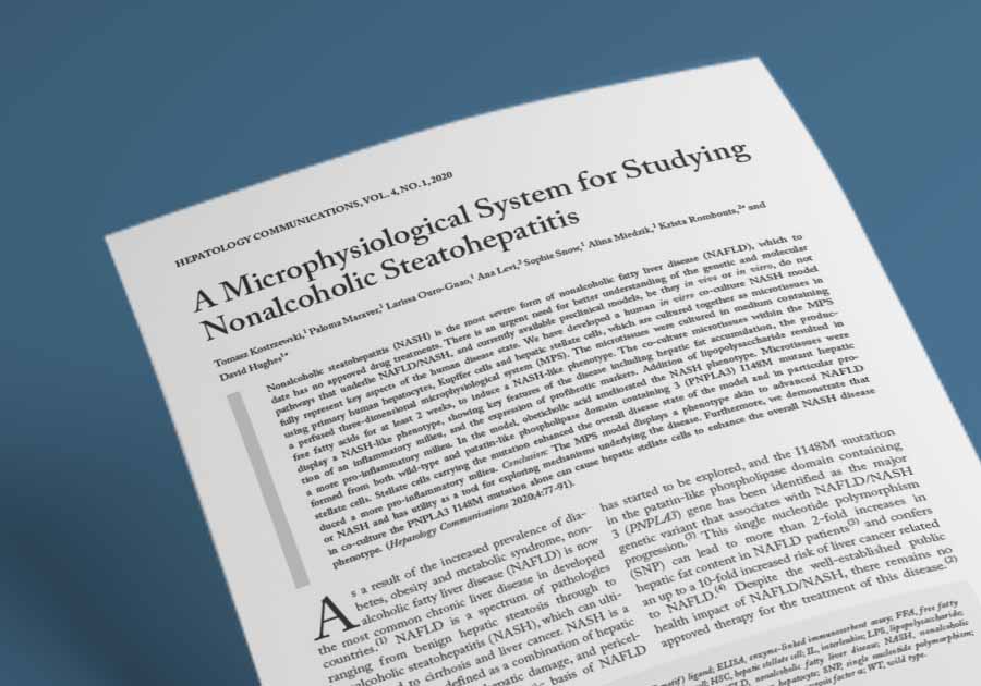
A Microphysiological System for Studying Non-alcoholic Steatohepatitis
Filed under: Disease modeling and MASLD/MASH
Summary
Researchers at CN Bio Innovations and University College London used CN Bio’s perfused 3D liver MPS, the platform now supplied as PhysioMimix Core with its liver-on-a-chip model, to build a human in vitro model of NASH from primary human hepatocytes, Kupffer cells and hepatic stellate cells held under high-fat conditions for 15 days. The co-culture microtissues accumulated intracellular fat, secreted pro-inflammatory cytokines and profibrotic markers, and carried a transcriptional profile distinct from a hepatocyte-only steatosis model run on the same platform. Obeticholic acid, an anti-NASH compound then in phase III trials, reduced IL-6 release and inflammatory gene expression in the model, and stellate cells carrying the PNPLA3 I148M variant produced a more pro-inflammatory and more steatotic phenotype than wild-type stellate cells. For drug discovery and disease-modeling teams, the study provides a human, donor-configurable NASH model with a two-week dosing window and a defined set of clinically recognized readouts. (NASH has since been renamed metabolic dysfunction-associated steatohepatitis, or MASH; this summary uses the terminology of the publication.)
Study facts at a glance
| Publication | Kostrzewski T, Maraver P, Ouro-Gnao L, Levi A, Snow S, Miedzik A, Rombouts K, Hughes D. A Microphysiological System for Studying Nonalcoholic Steatohepatitis. Hepatology Communications. January 2020;4(1):77-91. |
| DOI | 10.1002/hep4.1450 |
| CN Bio product used | PhysioMimix® Multi-chip Liver-12 plate and PhysioMimix Core System |
| How the platform was used | Primary human hepatocytes (PHH) were seeded at 6 x 10^5 viable cells per well, either alone or in co-culture with primary human Kupffer cells (HK) and primary human hepatic stellate cells (HSC) at 6×10⁴ viable cells each (approximately 10 hepatocytes to every Kupffer cell and stellate cell), in 1.6 mL of medium on collagen-coated 3D scaffolds, then perfused at a flow rate of 1.0 µL/s for 15 days in high free fatty acid (FFA) medium to induce a nonalcoholic steatohepatitis (NASH) phenotype. |
| Biological context | Human liver, nonalcoholic fatty liver disease (NAFLD) and its severe form NASH. All-human primary tri-culture of PHH, HK and HSC, with HSCs isolated from resected liver wedges or from livers unsuitable for transplant. Nine HSC donors were used across the study, including donors genotyped as homozygous wild type (WT) and homozygous I148M mutant for patatin-like phospholipase domain containing 3 (PNPLA3). The disease state was induced in vitro by fat loading rather than sourced from patients with diagnosed NASH. |
| Comparator | The PHH-only steatosis model previously developed in the same MPS, PHHs cultured in the absence of fat loading, lean (HEP-LEAN) medium controls, dimethyl sulfoxide (DMSO) vehicle controls for obeticholic acid (OCA) dosing, and WT versus I148M PNPLA3 mutant HSC co-cultures. |
| Key readouts | Intracellular fat by Oil Red O staining and FFA consumption from medium; interleukin 6 (IL-6) and tumor necrosis factor α (TNF-α) by enzyme-linked immunosorbent assay (ELISA); the fibrosis markers tissue inhibitor of metalloproteinase 1 (TIMP-1), fibronectin and procollagen 1 by ELISA; albumin secretion; a 27-plex Luminex cytokine, chemokine and growth factor panel combined with chemokine (C-X-C motif) ligand 1 (CXCL1); gene expression by RT2 Profiler PCR arrays covering 84 fatty liver genes and a cytokine and chemokine panel, plus Taqman quantitative PCR; PNPLA3 rs738409 genotyping. |
| Main interpretation | A perfused liver MPS maintained a PHH, HK and HSC co-culture for 15 days under high-fat conditions and produced a phenotype akin to advanced NAFLD or NASH, covering fat accumulation, an inflammatory milieu, profibrotic marker expression, a distinct transcriptional profile and a measurable response to OCA, and it showed that the PNPLA3 I148M variant in stellate cells alone can intensify that phenotype. |
Table of Contents
Which CN Bio product was used?
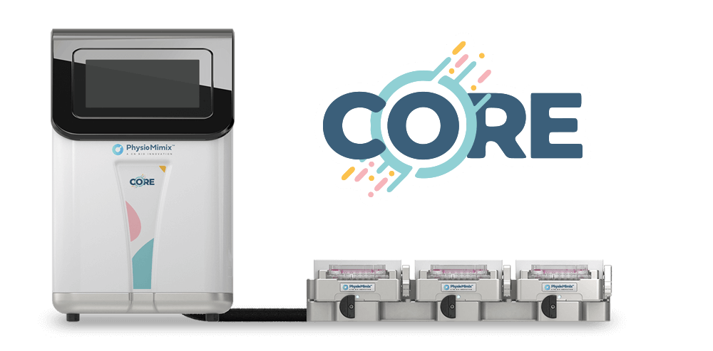
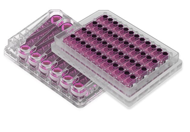
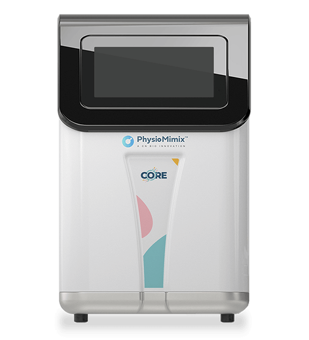
The study used the CN Bio LiverChip, the perfused 3D liver MPS that CN Bio now supplies as the PhysioMimix Core system. Cells were seeded onto collagen-coated scaffolds in 1.6 mL of medium per well and maintained under continuous perfusion at 1.0 µL/s, with PHHs at 6 x 10^5 viable cells and HKs and HSCs at 6 x 10^4 viable cells each, giving the approximate physiological ratio of 10 hepatocytes to every Kupffer cell and stellate cell. Cells were seeded in Williams E medium with primary hepatocyte thawing and plating supplements for the first 24 hours, then switched to HEP-LEAN or HEP-FAT medium, the CN Bio proprietary media that are now supplied in the NASH-in-a-box kit. HEP-FAT is a derivative of HEP-LEAN containing a mixture of saturated and unsaturated FFAs alongside physiologically relevant quantities of insulin and sugars. Complete medium changes were performed every 48 to 72 hours, and cultures ran for 15 days.
The platform carried every cell culture and compound dosing experiment in the paper, including fat loading, chronic low-dose lipopolysaccharide (LPS) challenge, OCA dosing and the wild-type versus mutant stellate cell comparison. Analytical work was performed off-platform on medium and on RNA recovered from the microtissues, using ELISA, Luminex, Oil Red O staining, RT2 Profiler arrays and Taqman quantitative PCR.
Find out more about CN Bio Liver-on-a-chip models here
Find out more about CN Bio DILI assays here
What this paper is about
NAFLD is the most common chronic liver disease in developed countries, and NASH is its severe form, defined by hepatic steatosis, inflammation, hepatic damage and pericellular fibrosis. At the time of publication there was no approved therapy. Preclinical progress had rested largely on rodent models, of which more than 30 had been developed without consensus on which is most useful, and on short in vitro exposures of 48 to 72 hours that capture transient responses to a triglyceride challenge rather than a chronic disease state. Every model type has strengths and limitations, and the specific gap this study addresses is duration and cellular composition: a human model that holds primary liver cells in a disease-inducing environment long enough for inflammation, profibrotic signaling and compound response to develop.
The approach was to move from a hepatocyte-only steatosis model, previously developed on the same platform, to a tri-culture that adds the two nonparenchymal populations most associated with initiating NASH: Kupffer cells, which drive the inflammatory response, and hepatic stellate cells, which drive fibrosis. This is the tri-culture architecture that underpins CN Bio’s current steatohepatitis disease-modeling assay. The design is modular: each cell type is added individually, so cells with a defined genetic background can be substituted into an otherwise fixed model, which is how the PNPLA3 I148M arm of the study was constructed.
What the researchers found
The tri-culture NASH model separated from the hepatocyte-only steatosis model on inflammation, fibrosis markers and transcriptional profile, but not on fat. The study reported IL-6 and TNF-α secretion, a range of upregulated pro-inflammatory genes, and higher concentrations of TIMP-1, fibronectin and procollagen 1 in the culture medium, alongside significant upregulation of the profibrotic genes CYGB, ACTA2, PCDH7, DES, COL1A1 and COL3A1. Both models accumulated fat at a similar rate and reached equivalent intracellular fat loading, and neither showed signs of hepatotoxicity. Albumin secretion was slightly lower in the NASH model, which matches the clinical observation that albumin falls in advanced liver disease. Of 84 fatty liver genes profiled against PHHs cultured without fat loading, 25 were differentially expressed in the NASH model compared with 6 in the steatosis model, with changes spanning insulin signaling, glucose metabolism, cholesterol metabolism, lipid metabolism and inflammation. CN Bio has separately compared this model’s profile against murine models and patient liver tissue in this resource.
Fat and endotoxin acted together on the inflammatory state. Repeat low-dose LPS from Escherichia coli O111:B4, added at 0.5 ng/mL from day 8 onward to mimic the gut-derived endotoxin exposure seen in patients, raised IL-6 and TNF-α further, while fat loading alone raised IL-6. Neither fat nor LPS altered procollagen 1 secretion, and hepatocytes in fat and fat plus LPS conditions produced less albumin than those in lean medium.
The model responded to pharmacological intervention. OCA, dosed from day 8 for a further week at 0.5 µM (approximating the plasma maximum concentration in patients) and 5 µM (simulating localized liver exposure), significantly reduced IL-6 release relative to vehicle controls, and reduced expression of inflammatory genes including monocyte chemoattractant protein 1 (MCP1), interferon-inducible protein 10 (IP-10) and TNF-α, plus the fibrosis-associated genes transforming growth factor β (TGF-β) and ACTA2. Genes associated with cholesterol metabolism, including CYP7A1, were downregulated and several glucose metabolism genes were upregulated. The authors noted a significant fall in IL-6 protein with only minimal change in IL-6 gene expression, suggesting an effect on translation, post-translational modification or secretion.
Genetic background changed the disease phenotype. Co-cultures built with HSCs homozygous for the I148M PNPLA3 variant produced significantly more IL-6 than those with wild-type HSCs at both day 8 and day 15, with the highest levels in LPS-dosed samples, and also showed reduced albumin secretion, greater fat accumulation and further upregulation of ACTA2, IGFBP1, IL-6 and GK. Luminex profiling showed increased secretion of cytokines associated with chronic inflammation, including interferon γ, IL-6 and IL-12, and of growth factors associated with tissue remodeling and stellate cell activation, including platelet-derived growth factor and MCP1. The variant did not alter secretion of the profibrotic markers procollagen 1 and TIMP-1.
Why the paper matters
For teams selecting a preclinical model for metabolic liver disease, the practical contribution is a human model with a working duration that suits repeat compound dosing. The co-cultures were maintained in a functional state for at least two weeks, which allowed an eight-day disease induction phase followed by a seven-day dosing phase within a single experiment, and allowed longitudinal sampling of the same cultures at days 8, 11, 13 and 15. The paper positions this as a step towards the time scale of rodent models rather than a replacement for them, and reports the model as cost-comparable to genetic and diet-induced rodent NAFLD models, with components to generate each 3D co-culture microtissue costing US $150 to $400 depending on cell source. Groups that would rather commission the work than run it in-house can access the same model through CN Bio’s MASH in vitro services.
The second contribution is mechanistic. Because each cell type is added separately, the model can isolate the contribution of one population’s genotype while holding the rest of the system constant. That design produced a finding that monoculture work could not: the PNPLA3 I148M variant in stellate cells alone was sufficient to enhance the overall NASH phenotype of the co-culture, and its effects were most pronounced under LPS challenge, pointing to a possible combinatorial effect between the variant and gut-derived endotoxin as two risk factors in patients. The same modular logic applies to other organ-on-a-chip models in which specific genetic backgrounds or donor characteristics are the experimental variable.
Key study takeaways
- The study used the CN Bio perfused 3D liver MPS, named LiverChip in the publication and now supplied as PhysioMimix Core, to co-culture primary human hepatocytes, Kupffer cells and hepatic stellate cells for 15 days under high free fatty acid conditions.
- The model reproduced key features of NASH, including intracellular fat accumulation, secretion of IL-6 and TNF-α, and production of the profibrotic markers TIMP-1, fibronectin and procollagen 1.
- Compared with the hepatocyte-only steatosis model on the same platform, the tri-culture NASH model showed higher inflammation and fibrosis markers and a broader transcriptional shift (25 of 84 fatty liver genes differentially expressed versus 6), with equivalent fat loading in both.
- The workflow combined ELISA biomarker measurement, Luminex multiplex cytokine profiling, Oil Red O fat quantification, targeted and array-based gene expression, and PNPLA3 genotyping of the stellate cell donors.
- OCA reduced IL-6 secretion and inflammatory gene expression in the model, supporting use of the platform for screening anti-NASH compounds and investigating their mechanism of action.
- The model is most appropriate for mechanistic and compound-response questions over a two-week window. It does not include liver sinusoidal endothelial cells or peripheral immune cells, does not recapitulate full hepatic zonation, and compresses a disease that develops over decades in patients into 15 days, so results should be read with that temporal difference in mind.
Why this paper is worth reading
For anyone selecting a liver model, the publication is one of the few places where eight commercial liver MPS are described side by side, including details on the cell types, configuration, format, donor numbers, cell numbers per chip, exposure volumes, culture duration, metabolic competence, hepatocyte biomarkers, bile canaliculi and transport, and the published hepatotoxicity prediction datasets behind each system. That makes it a useful reference for scoping a safety toxicology study, whatever platform a team ends up choosing.
For regulatory strategy, the value sits in the framing. The project treats a liver MPS as a weight-of-evidence tool tied to one clinical question rather than a general-purpose replacement for animal testing, and it builds performance expectations at the level of the model category. Liver MPS results should not be read as a stand-alone binary classifier, partly because the low clinical prevalence of DILI, estimated at roughly 13.9 to 19.1 cases per 100,000 individuals annually, limits positive predictive value even for assays with high sensitivity and specificity.
FAQ
The study used the CN Bio LiverChip, a perfused three-dimensional liver microphysiological system from CN Bio Innovations Ltd. This is the liver platform CN Bio now supplies as the PhysioMimix Core system, run with the HEP-LEAN and HEP-FAT media now provided in the NASH-in-a-box kit.
The LiverChip MPS held primary human hepatocytes, Kupffer cells and hepatic stellate cells as 3D microtissues on collagen-coated scaffolds in 1.6 mL of medium per well, under perfusion at 1.0 µL/s for 15 days. Fat loading with HEP-FAT medium began on day 1, LPS dosing at 0.5 ng/mL began on day 8, and OCA was dosed from day 8 for a further seven days.
The paper models nonalcoholic steatohepatitis, the severe form of nonalcoholic fatty liver disease, in an all-human primary liver co-culture of hepatocytes, Kupffer cells and hepatic stellate cells. Stellate cells were genotyped for the PNPLA3 I148M variant, and both wild-type and homozygous mutant donors were used.
The tri-culture liver MPS produced a phenotype akin to advanced NAFLD or NASH, with hepatic fat accumulation, an inflammatory milieu and profibrotic marker expression, and it responded to obeticholic acid with reduced IL-6 release. The study also showed that hepatic stellate cells carrying the PNPLA3 I148M variant alone enhanced the overall NASH phenotype, particularly under LPS challenge.
The main comparator was the hepatocyte-only steatosis model previously developed on the same MPS, along with PHHs cultured without fat loading, lean medium controls, DMSO vehicle controls for OCA dosing, and wild-type versus I148M PNPLA3 mutant hepatic stellate cell co-cultures.
Readouts included Oil Red O fat quantification and FFA consumption, ELISA measurement of IL-6, TNF-α, TIMP-1, fibronectin, procollagen 1 and albumin, a 27-plex Luminex cytokine and chemokine panel with CXCL1, RT2 Profiler and Taqman gene expression analysis, and PNPLA3 rs738409 genotyping.
The paper gives drug discovery and disease-modeling teams a fully specified human NASH model with a two-week dosing window, clinically recognized biomarker readouts and a documented pharmacological response to obeticholic acid. It is equally useful for its stated limits: the model excludes liver sinusoidal endothelial cells and peripheral immune cells, does not recapitulate hepatic zonation, and compresses a decades-long human disease into 15 days of culture.
Full citation
Related products and services
Contract research services
Discover how to utilize our cross-species models to inform next-step decision making via our DILI in vitro Contract Research Services here.
Add PhysioMimix Core in your lab
To develop your own cross-species Liver MPS models, you will need: