
Resource > Videos and animations >
Lung-on-a-chip Technology: How does it work?
Mimicking the alveolar and a bronchial airway
Filed under: Drug absorption and Pulmonary infection
To map the distinct areas and physiology of the human lung, we created two Innovate UK grant funded Lung on a chip models that mimic the alveolar and a bronchial airway.
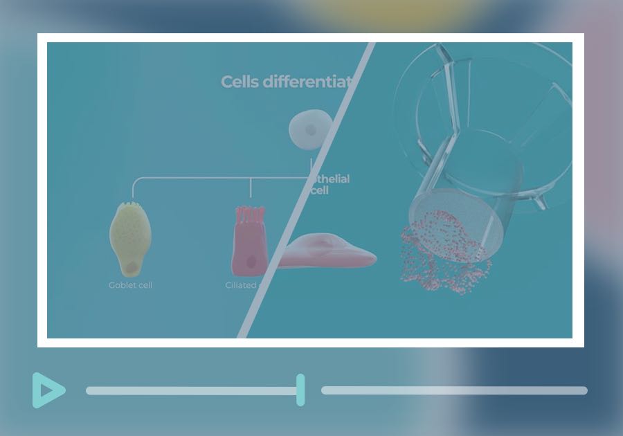
Our animations provide a visual representation of how Lung on a chip technology works.
Human alveolar microphysiological system
Human bronchial microphysiological system
Dynamically perfused Lung on a chip technology is constructed using the PhysioMimix® Core microphysiological system and Multi-chip Barrier plates.
Primary human small airway or bronchial epithelial cells are cultured for 14 days at air liquid interface (ALI) on Transwell® inserts in PhysioMimix Multi-chip Barrier plates.
Enhanced physiological relevance (versus static 2D cultures) is incorporated into Lung-on-a-chip models, also known as Lung microphysiological systems (Lung MPS), by including primary human pulmonary microvascular endothelial cells on the basolateral side of a perfused Transwell®.
Monocytes are also incorporated, either basolaterally in the bronchial model to represent circulating immune cells, or on the apical side of the alveolar model to act as an alveolar macrophage-like cell.
Validation studies demonstrate that perfused multi-cell type lung-on-a-chip technology accurately mimics human lung tissue, demonstrating relevant tissue architectures, cellular differentiation (diversity) and function.
The advantages of lung on a chip technology are described in more detail on our dedicated web page, however, the technology has been proven to enhance the predictive power and clinical relevance of airway models for in vitro studies (Caygill et al., 2025, Alveolar and bronchial microphysiological systems for respiratory infection research and therapeutics evaluation Application Note, Phan et al., 2023).