

Resource > Scientific publications >
Modelling human liver fibrosis in the context of non-alcoholic steatohepatitis using a microphysiological system
Filed under: Disease modeling and MASLD/MASH
Summary
The PhysioMimix® LC12 MPS was used to build a tri-culture human liver model of metabolic dysfunction-associated steatohepatitis (MASH) that produces a measurable fibrosis phenotype. Primary human hepatocytes were co-cultured with primary human Kupffer cells and hepatic stellate cells under continuous perfusion, then exposed to free fatty acids and selected biological cues, which generated collagen-1 deposition, alpha-smooth muscle actin (α-SMA) expression, and a gene expression signature that matched human MASH patient samples much more closely than standard rodent models. The model responded to the clinical-stage compounds obeticholic acid (OCA) and elafibranor (ELF) and to a dietary-style switch from high-fat to lean medium, which makes it suitable for studying MASH biology and for testing anti-inflammatory and anti-fibrotic interventions across a range of disease severity.
Study facts at a glance
| Publication | Kostrzewski T, Snow S, Lindstrom Battle A, Peel S, Ahmad Z, Basak J, Surakala M, Bornot A, Lindgren J, Ryaboshapkina M, Clausen M, Linden D, Maass C, Young LM, Corrigan A, Ewart L, Hughes D. Modelling human liver fibrosis in the context of non-alcoholic steatohepatitis using a microphysiological system. Communications Biology. 2021;4:1080. |
| DOI | 10.1038/s42003-021-02616-x |
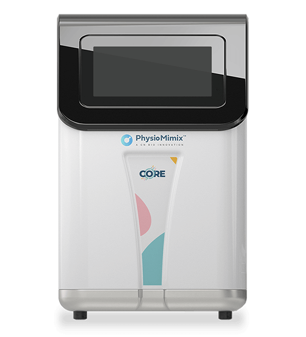
| CN Bio product used | PhysioMimix® LC12 and PhysioMimix Core System |
| How the platform was used | A tri-culture of primary human hepatocytes, Kupffer cells, and hepatic stellate cells was cultured under continuous perfusion (1.0 microL/s) for 14 to 30 days, with free fatty acids and biological cues (TGFβ, LPS, fructose, cholesterol) added to model steatosis, inflammation, and fibrosis |
| Biological context | Human metabolic dysfunction-associated steatohepatitis (MASH) within the metabolic dysfunction–associated steatotic liver disease (MASLD) spectrum; primary human liver cells; conditions spanning lean control, steatosis, and MASH |
| Comparator | Control and steatotic microtissues, published human MASH patient transcriptomic datasets, nine standard mouse MASH models, and vehicle controls for drug studies |
| Key readouts | Quantitative confocal imaging of collagen-1 and alpha-smooth muscle actin (α-SMA); secreted fibrosis biomarkers and cytokines by ELISA and Luminex; gene expression by RNA sequencing and PCR arrays; albumin secretion; Oil Red O fat staining; drug half-life and protein binding by LC-MS/MS |
| Main interpretation | The model generates a quantifiable fibrosis phenotype and a transcriptomic profile closer to human MASH than common rodent models, supporting its use for MASH mechanistic studies and for testing anti-fibrotic and anti-inflammatory drug candidates across disease stages |
Table of Contents
Which CN Bio product was used?
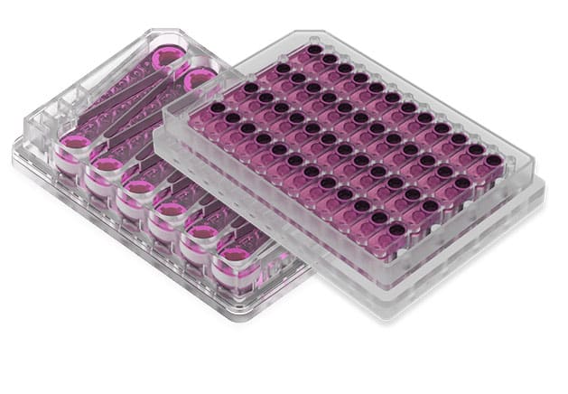
The study used the PhysioMimix LC12 MPS from CN Bio Innovations. Each LC12 plate holds 12 individual bioreactors, and each bioreactor contains a pneumatically driven micro-pump that recirculates cell culture medium through a collagen-coated scaffold, giving every liver microtissue continuous perfusion for the length of the experiment. Primary human hepatocytes were seeded at 6 x 10^5 cells per well, with Kupffer cells and hepatic stellate cells added at either a 1:10 ratio (high non-parenchymal cell content) or a 1:100 ratio (low non-parenchymal cell content). Cultures ran at a flow rate of 1.0 microL/s in proprietary HEP-Lean or HEP-Fat media, with the high-fat medium supplying saturated and unsaturated free fatty acids alongside physiological levels of insulin and sugars. Standard experiments lasted 14 days, reversibility studies were extended to 30 days, and viability was maintained for at least 4 weeks.
The PhysioMimix platform was central to the work, because it generated and maintained the perfused liver microtissues used in every condition. Downstream characterization, including confocal imaging, ELISA, Luminex cytokine arrays, RNA sequencing, and LC-MS/MS drug analysis, was carried out on the microtissues and culture media produced in the platform.
What this paper is about
MASH is a progressive form of MASLD marked by fat accumulation in hepatocytes, immune cell infiltration, hepatocellular ballooning, collagen deposition, and liver fibrosis. There are no approved NASH therapies, partly because existing laboratory models do not reproduce the disease well. Most in vitro models lack a strong, measurable fibrosis signal, and rodent models often fail to mirror the gene expression patterns seen in human patients. Fibrosis severity is the main predictor of progression to cirrhosis and of liver-related mortality in MASLD, so a model that captures it is valuable for drug discovery.
This paper develops and characterizes a human liver MASH model built in the PhysioMimix MPS. Hepatocytes, Kupffer cells, and hepatic stellate cells were co-cultured under perfusion to recreate the multicellular liver microenvironment. The authors added an automated confocal imaging assay to quantify collagen-1 deposition and alpha-smooth muscle actin (α-SMA), ran full transcriptomic and biomarker profiling, compared the results against human patient datasets and rodent models, and tested both drug treatment and dietary-style intervention. A systems biology approach was then used to identify which biological cues push the model toward advanced disease.
What the researchers found
The MASH tri-culture produced a clear, quantifiable fibrosis phenotype. Compared with control and steatotic microtissues, MASH microtissues showed a sixfold increase in alpha-smooth muscle actin (α-SMA) expression and an eightfold increase in collagen-1 deposition, measured by automated confocal imaging. These changes were backed up by significant increases in secreted clinical fibrosis biomarkers (TIMP-1, pro-collagen 1, YKL-40, and fibronectin) and by raised expression of fibrosis-associated genes.
The gene expression profile of the model aligned with human disease. Transcriptomic data from the MASH microtissues mapped to a human liver and human fatty liver disease signature, and 77% of the top 100 expressed genes related to hepatic function, metabolism, inflammation, and fibrosis. When compared with published patient datasets, up to 45% of the differentially expressed genes from MASH patients were also correctly changed in the model. The same patient gene sets were reflected in only 0.01% to 10% of cases across nine standard mouse MASH models, which places the MPS model much closer to human disease.
The model responded to drugs in a dose-dependent way. Obeticholic acid (OCA), a farnesoid X receptor (FXR) agonist, and elafibranor (ELF), a dual PPARα/δ agonist, both lowered inflammatory cytokines and reduced expression of genes tied to extracellular matrix remodeling (MMP1, MMP9, SERPINA1), TGFβ signaling (CEBPB, DCN), and inflammation (CCL2, CCL3, CXCR4). In the fat-only MASH model, the two compounds cut α-SMA expression by up to 50% and collagen-1 deposition by up to 35%. Both drugs bound strongly to protein in the culture medium and showed measured half-lives of 12 hours (OCA) and 16 hours (ELF) in the metabolically active microtissues, which the authors used to set clinically relevant dosing.
Disease markers were reversible. Switching MASH microtissues from high-fat to lean medium partway through a 30-day culture reduced inflammation (IL-6 and MCP-1), lowered the fibrosis marker TIMP-1, and halted further fat loading, mimicking the effect of dietary change in patients.
Specific cues drove a more advanced disease state. Using a systems biology cue-signal-analysis approach, the team tested free fatty acids, lipopolysaccharide (LPS), transforming growth factor beta (TGFβ), fructose, and cholesterol alone and in combination. Free fatty acids, with or without cholesterol, modeled steatosis, while TGFβ, and to a lesser extent fructose, drove inflammation, fibrosis, and reduced liver function consistent with advanced MASH. In this TGFβ-supplemented model, OCA and ELF produced even larger effects, with the highest doses reducing α-SMA by 80% and collagen-1 by 75%.
Why the paper matters
For drug discovery teams working on MASH, the value of this model is a human-relevant fibrosis readout that can be measured and reproduced. Fibrosis is the histological feature most closely tied to poor outcomes in MASLD and the hardest to reproduce in rodents, so an in vitro model that generates quantifiable collagen-1 and alpha-smooth muscle actin (α-SMA) signals gives teams a way to test anti-fibrotic candidates directly on human cells. Because the transcriptomic profile sits closer to human patients than common mouse models, results carry more translational weight when selecting compounds or studying disease pathways.
The model also covers a spectrum of disease. The same platform can represent simple steatosis, mid-stage MASH, and advanced MASH with marked fibrosis, depending on the cues applied, which lets one system test a compound across several disease states, potentially in parallel. Drug responses can be read in about two weeks, compared with months in rodent studies, and the authors scaled in vitro doses to clinically relevant exposures, an approach they recommend for other advanced in vitro work. The perfused, multicellular format keeps primary human cells viable and functional for at least four weeks, which supports chronic and repeat-dose studies that are closer to in vivo conditions while reducing reliance on animals.
Key study takeaways
- The study used the PhysioMimix LC12 MPS to build a perfused tri-culture of primary human hepatocytes, Kupffer cells, and hepatic stellate cells that models MASH.
- The model reproduced a measurable fibrosis phenotype, with a sixfold rise in α-SMA and an eightfold rise in collagen-1 deposition in MASH microtissues versus controls.
- Compared with standard mouse MASH models, the model matched human patient gene expression far more closely, recapitulating up to 45% of patient differentially expressed genes versus 0.01% to 10% for rodent models.
- The workflow combined automated confocal imaging of collagen-1 and α-SMA, secreted biomarker measurement (TIMP-1, pro-collagen 1, YKL-40, fibronectin), Luminex cytokine arrays, and RNA sequencing.
- The findings support using the liver MPS MASH model to test anti-inflammatory and anti-fibrotic compounds, shown here with obeticholic acid (OCA) and elafibranor (ELF), as well as dietary-style intervention.
- The model is most useful when matched to a defined disease stage: free fatty acids with or without cholesterol for steatosis, and transforming growth factor beta (TGFβ) with free fatty acids for advanced MASH with fibrosis. Adding extra-hepatic stimuli would be needed to push the model toward cirrhosis.
Why this paper is worth reading
This paper is useful because it gives drug discovery and disease-modeling scientists a documented way to generate and quantify liver fibrosis in a human in vitro system, which has been one of the weak points of earlier MASH models. It sets out the cell ratios, perfusion conditions, biological cues, imaging assay, and dosing logic needed to model disease from steatosis through to advanced MASH, and it benchmarks the model against both human patient data and rodent models so readers can judge its translational reach. For teams deciding which preclinical model to use, or designing compound efficacy studies, it offers a practical framework and a clear set of measurable endpoints rather than a single isolated result.
FAQs
The study used the PhysioMimix LC12 MPS from CN Bio Innovations, running tri-culture liver microtissues in LC12 plates with collagen-coated, perfused bioreactors.
The PhysioMimix LC12 MPS maintained primary human hepatocytes, Kupffer cells, and hepatic stellate cells under continuous perfusion at 1.0 microL/s for 14 to 30 days, with free fatty acids and cues such as TGFβ, LPS, fructose, and cholesterol added to model steatosis, inflammation, and fibrosis.
The paper studied MASH, a severe form of MASLD, using a human liver tri-culture of primary hepatocytes, Kupffer cells, and hepatic stellate cells.
The main finding was that the liver MPS MASH model produces a quantifiable fibrosis phenotype, with large increases in collagen-1 deposition and alpha-smooth muscle actin (α-SMA), and a transcriptomic profile that matches human MASH patients much more closely than standard rodent models.
The study compared MASH microtissues against control and steatotic microtissues, against published human MASH patient gene expression datasets, and against nine standard mouse MASH models, and it compared drug-treated microtissues against vehicle controls.
Readouts included automated confocal imaging of collagen-1 and α-SMA, secreted fibrosis biomarkers (TIMP-1, pro-collagen 1, YKL-40, fibronectin) and cytokines by ELISA and Luminex, gene expression by RNA sequencing and PCR arrays, albumin secretion, Oil Red O fat staining, and drug half-life and protein binding by LC-MS/MS.
The paper is useful because it provides a human-relevant liver MPS MASH model with a measurable fibrosis endpoint, suited to testing anti-fibrotic and anti-inflammatory compounds and to studying MASH disease mechanisms across a spectrum of severity.
Full citation
Related products and services
Contract research services
Discover how to utilize our cross-species models to inform next-step decision making via our DILI in vitro Contract Research Services here.
Add PhysioMimix Core in your lab
To develop your own cross-species Liver MPS models, you will need: