




Resource > Scientific publications >
Human Liver MPS for Assessing Drug-Induced Liver Toxicity In Vitro
Filed under: DILI and Safety toxicology
Summary
This study used the PhysioMimix® system and Multi-chip Liver-12 plates to build perfused 3D liver microtissues from cocultured primary human hepatocytes (PHHs) and human Kupffer cells (HKCs), then tested whether the model could predict drug-induced liver injury (DILI). Dosing two structurally similar compounds at seven concentrations for up to 96 hours, the liver MPS flagged severe toxicity for troglitazone (rising ALT and LDH, with sharp falls in albumin and urea) while registering only mild effects for pioglitazone, matching each drug’s known clinical DILI risk. The work matters for drug discovery and toxicology because the liver MPS detected a hepatotoxicant that standard 2D and some 3D liver models miss, and it did so reproducibly using clinically relevant readouts such as ALT.
Study facts at a glance
| Publication | Novac O, Silva R, Young L-M, Lachani K, Hughes D, Kostrzewski T. Human Liver Microphysiological System for Assessing Drug-Induced Liver Toxicity In Vitro. J Vis Exp (JoVE). 2022 Jan 31;(179):e63389. |
| DOI | 10.3791/63389 |
| CN Bio product used | PhysioMimix system with the Multi-chip Liver-12 plate (MPS-LC12). |
| How the platform was used | Cryopreserved PHHs and human Kupffer cells HKCs were cocultured on the scaffolds of the Multi-chip Liver-12 plate and perfused under flow to form 3D liver microtissues, then taken through an 8-day DILI assay with quality control at Day 4 and compound dosing every 48 hours across seven test concentrations for up to 96 hours. |
| Biological context | DILI in human liver. The model used 3D liver microtissues built from cocultured PHHs and HKCs (the resident liver macrophages and a non-parenchymal cell type), derived from cryopreserved donor primary cells in a healthy (non-diseased) liver context. |
| Comparator | Within the study, treated microtissues were measured against vehicle controls and a chlorpromazine positive control (100 µM). The paper also contrasts the liver MPS with standard 2D hepatocyte cultures and some 3D models that had failed to detect troglitazone toxicity. |
| Key readouts | ALT release, LDH release, albumin production, urea synthesis, CYP3A4 activity, ATP content, and brightfield and IF microscopy. LDH and urea were also used for Day 4 quality control. |
| Main interpretation | The liver MPS reproducibly separated a severe hepatotoxicant (troglitazone) from a compound of low DILI concern (pioglitazone) across several functional endpoints, supporting its use for mechanistic DILI assessment in preclinical drug development. |
Table of Contents
- Summary
- Study facts at a glance
- Which CN Bio product was used?
- What this paper is about
- What the researchers found
- Why the paper matters
- Key study takeaways
- Why this paper is worth reading
- FAQs
- Full citation
Which CN Bio product was used?
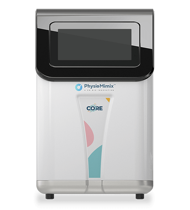
The study used the PhysioMimix system with the Multi-chip Liver-12 plate. The platform paired a controller and a pneumatic docking station with an MPS driver that holds the consumable plate. Experiments were ran on the Multi-chip Liver-12 (LC12) plate, which has 12 independent wells, each containing a collagen-coated scaffold with microchannels. Embedded micropumps circulate culture medium through the scaffolds under flow, so the primary cells form and maintain 3D liver microtissues rather than flat monolayers.
In this work the platform carried the full assay: priming and quality control, microtissue formation, repeat compound dosing, and endpoint sampling. This system has been sown to keep liver microtissues functional under perfusion for up to 4 weeks, and it has been evaluated by the US Food and Drug Administration (FDA) with high reproducibility for drug toxicity, metabolism, and intracellular accumulation studies, including higher sensitivity than spheroid and sandwich cultures for several drugs.
What this paper is about
DILI is a leading cause of acute liver failure and a major reason drug candidates fail in development. More than 1,000 FDA-approved medicines are known to carry DILI potential, and the injury often goes undetected until a drug reaches clinical stages, putting patients at risk and creating large losses for developers. The problem is partly a model problem. Standard 2D hepatic cell lines such as HepG2 and HepaRG come from single donors and show abnormal liver function, while plated PHHs lose hepatic function quickly and have low sensitivity (under 50%) for DILI prediction. Animal testing is slow, costly, requires cross-species translation, and suits human-specific drug modalities poorly.
A human liver MPS, a type of organ-on-a-chip (OOC), was used to address these gaps. Perfused 3D microtissues were formed from PHHs cocultured with HKCs, and the the functional read outs included clinically relevant biomarkers to allow comparison against in vivo and clinical observations. To test the model, the team compared two structurally similar compounds with opposite clinical DILI profiles: troglitazone, a type 2 diabetes drug withdrawn from the market over liver injury, and pioglitazone, a compound of low DILI concern.
Learn more about our DILI-related offerings
What the researchers found
The liver MPS produced consistent microtissues and reproducible data. Across three experiments, control microtissues kept high albumin and urea output, CYP3A4 activity, and ATP content over the 8-day culture, with low intra-study and inter-study variability.
For troglitazone, the model showed an acute, peak-plasma-concentration-driven (Cmax-driven) toxic response. After 96 hours of exposure, the study reported ALT and LDH release together with rapid drops in albumin and urea production at around 15 times the clinical Cmax. ATP content and CYP3A4 activity confirmed the toxicity, and the half-maximal effective concentration (EC50) values agreed closely across endpoints. Brightfield microscopy showed generalised microtissue death at the top two test concentrations, against healthy, evenly seeded vehicle-control microtissues.
For pioglitazone, the model registered only mild effects, in line with its low clinical DILI concern. No ALT or LDH release was detected. After 48 hours, the study reported a small reduction in albumin and urea production at roughly 25 times the clinical Cmax, with a minor, non-significant drop in ATP content at high concentrations and slight microtissue alteration at the two highest concentrations by 96 hours.
Combining six functional endpoints (ALT release, LDH release, albumin production, urea synthesis, CYP3A4 activity, and ATP content) across the 48-hour and 96-hour timepoints produced a heatmap “signature of hepatotoxicity” that separated the two compounds by DILI risk.
Why the paper matters
The result that matters most for safety teams is sensitivity with specificity. The liver MPS detected severe toxicity for troglitazone, a compound that published animal studies and standard 2D hepatic assays had failed to flag, and it graded pioglitazone as only mildly toxic. The model therefore tracked clinical DILI severity rather than simply reporting generic cytotoxicity.
Two further points support adoption. First, the endpoints are clinically measured biomarkers such as ALT, which makes the data easier to relate to in vivo and clinical findings. Second, the reproducibility shown here speaks to a known barrier to MPS uptake in industry: the lack of process standardisation and inter-site reproducibility that has kept MPS data out of routine screening and regulatory submissions. By pairing a worked protocol with reproducible results, the paper supports earlier, mechanism-aware preclinical DILI decisions and offers an approach that can reduce reliance on animal testing. The authors also note the model can be applied to disease states such as viral hepatitis and fatty liver disease, and to specific patient subsets.
Key study takeaways
- The study used the PhysioMimix platform with the Multi-chip Liver-12 plate to form perfused 3D microtissues from cocultured PHHs and HKCs, dosed across seven concentrations over a 96-hour window.
- Control microtissues stayed functional for 8 days, with high albumin and urea output, CYP3A4 activity, and ATP content, and low intra-study and inter-study variability across three runs.
- The model classified troglitazone as a severe hepatotoxicant, with ALT and LDH release and falling albumin and urea at about 15 times the clinical Cmax, matching its history of causing liver failure.
- The same model graded pioglitazone as mildly toxic, with no ALT or LDH release and only small albumin and urea reductions at about 25 times the clinical Cmax, in line with its low DILI concern.
- A heatmap built from six functional endpoints (ALT release, LDH release, albumin production, urea synthesis, CYP3A4 activity, and ATP content) gave a “signature of hepatotoxicity” that distinguishes compounds by DILI risk.
- The authors position the assay for later-stage preclinical mechanistic DILI testing rather than high-throughput screening, and note it can be extended to diseased liver backgrounds and specific patient subsets.
Why this paper is worth reading
This paper is useful because it gives drug discovery and safety teams a worked, reproducible protocol for grading DILI risk in a human liver model, not only a claim that the model works. It sets out the full 8-day workflow, the day-by-day dosing and quality-control steps, and the panel of endpoints needed to separate a withdrawn hepatotoxicant from a low-concern drug. For anyone deciding whether an organ-on-a-chip liver model earns a place in preclinical safety testing, the side-by-side troglitazone and pioglitazone data, together with the endpoint heatmap, offer a concrete framework to judge that decision against.
FAQs
The study used the PhysioMimix system with the Multi-chip Liver-12 (LC12) consumable plate and collagen-coated 3D scaffolds.
The PhysioMimix platform formed perfused 3D liver microtissues from cocultured primary human hepatocytes (PHHs) and human Kupffer cells (HKCs), then ran an 8-day assay with quality control at Day 4 and compound dosing every 48 hours across seven concentrations for up to 96 hours.
The model addressed drug-induced liver injury (DILI) in human liver, using 3D microtissues built from cryopreserved primary human hepatocytes (PHHs) and human Kupffer cells (HKCs).
The liver MPS detected severe, Cmax-driven toxicity for troglitazone and only mild effects for pioglitazone, matching the two drugs’ known clinical DILI profiles.
Within the study, treated microtissues were compared against vehicle controls and a chlorpromazine positive control (100 µM). The paper also contrasts the liver MPS with standard 2D hepatocyte cultures and some 3D models that had failed to detect troglitazone toxicity.
The study measured ALT release, LDH release, albumin production, urea synthesis, CYP3A4 activity, and ATP content, alongside brightfield and immunofluorescence microscopy, with LDH and urea also used for Day 4 quality control.
It provides a reproducible protocol and endpoint panel for grading DILI liability in a human liver organ-on-a-chip model, supporting earlier mechanistic safety decisions in preclinical drug development and offering an approach that can reduce reliance on animal testing.