

Resource > Scientific publications >
Bone morphogenetic protein 8B promotes the progression of Non-Alcoholic Steatohepatitis
Filed under: Disease modeling and MASLD/MASH
Summary
Researchers used the CN Bio LiverChip liver microphysiological system (MPS) to test, in primary human liver cells, whether BMP8B contributes to NASH progression. In a 3D perfused tri-culture of primary human hepatocytes, Kupffer cells, and hepatic stellate cells challenged with free fatty acids to induce a NASH phenotype, recombinant BMP8 phosphorylated SMAD1 and SMAD3, activated proinflammatory and proliferative pathways, and increased secretion of ALT, MCP1, RANTES, and M-CSF while suppressing the anti-inflammatory cytokines IL-10 and IL-13. The human model mirrored most of the pathway changes seen in BMP8B knockout mice, which supports BMP8B as a mechanistically plausible target in the early, inflammatory phase of NASH and shows how a human liver MPS can be used to test a target hypothesis generated in rodents.
Study facts at a glance
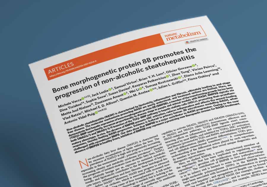
| Publication | Vacca M, Leslie J, Virtue S, Lam BYH, Govaere O, Tiniakos D, Snow S, Davies S, Petkevicius K, Tong Z, Peirce V, Nielsen MJ, Ament Z, Li W, Kostrzewski T, Leeming DJ, Ratziu V, Allison MED, Anstee QM, Griffin JL, Oakley F, Vidal-Puig A. Bone morphogenetic protein 8B promotes the progression of non-alcoholic steatohepatitis. Nature Metabolism. June 2020. 2(6):514-531. |
| DOI | 10.1038/s42255-020-0214-9 |
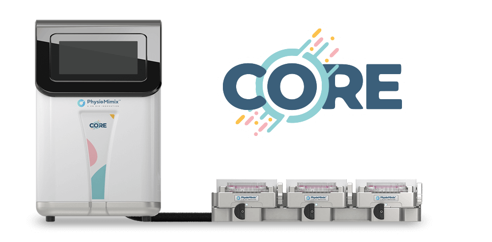
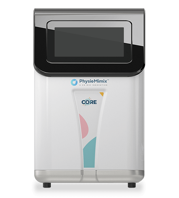
| CN Bio product used | PhysioMimix® LC12 and PhysioMimix Core System |
| How the platform was used | The liver MPS held 3D perfused microtissues of cryopreserved primary human hepatocytes, Kupffer cells, and hepatic stellate cells (HSCs) for one week in HEP-FAT medium, which contains a mixture of saturated and unsaturated free fatty acids (FFAs) with physiologically relevant quantities of insulin and sugars to induce a non-alcoholic steatohepatitis (NASH) phenotype. After 8 days, microtissues were treated for up to 48 hours with recombinant BMP8 (75 ng/mL), with or without an activin receptor-like kinase (ALK) 1/2/3/6 inhibitor (K02288, 1 µM), an ALK4/5/7 inhibitor (A-8301, 1 µM), transforming growth factor beta (TGFβ, 5 ng/mL), or BMP7 (5 ng/mL). Complete medium changes were performed every 48 hours. Flow rate and plate format are not specified in the paper. |
| Biological context | Liver, human NASH within non-alcoholic fatty liver disease (NAFLD) now referred to as metabolic dysfunction-associated steatotic liver disease (MASLD). All-human tri-culture of primary hepatocytes, Kupffer cells, and HSCs in a fatty acid induced disease state. The wider paper also covers two human NASH biopsy cohorts (n = 40 Cambridge; n = 113 Newcastle/Paris) and several mouse models of liver injury and NASH. |
| Comparator | Within the liver MPS: untreated FFA-challenged microtissues, with TGFβ and BMP7 as positive controls and the two ALK inhibitors as pathway specificity controls. Lean (non-FFA) medium served as the baseline for BMP8B expression. Across the wider paper: bone morphogenetic protein 8B (BMP8B) knockout mice versus wild-type littermates, plus a transcriptome-level comparison of the human in vitro NASH model against the western diet mouse NASH model. |
| Key readouts | RNA sequencing and quantitative real-time polymerase chain reaction (RTqPCR) of TGFβ-BMP target genes, inflammatory genes, proliferation drivers, and fibrosis markers; targeted phospho-proteomics of SMAD1 and SMAD3 and downstream kinases; secretome profiling of alanine aminotransferase (ALT), monocyte chemoattractant protein 1 (MCP1), RANTES, macrophage colony-stimulating factor (M-CSF), interleukin (IL) 10, IL-13, TIMP-1, and C3M; Oil Red O staining of lipid droplets; Ingenuity Pathway Analysis and upstream regulator analysis. |
| Main interpretation | The human liver MPS reproduced, in primary human cells, the BMP8-driven inflammatory and proliferative signaling that BMP8B knockout mice had implicated in NASH progression, while showing only mild and transient effects on stellate activation and fibrosis markers over 48 hours. |
Table of Contents
Which CN Bio product was used?
The study used the LiverChip MPS from CN Bio Innovations for its human in vitro arm. The model is an all-human, 3D perfused liver tri-culture: cryopreserved primary human hepatocytes, Kupffer cells, and hepatic stellate cells, cultured together under flow. Disease induction came from HEP-FAT medium, CN Bio’s proprietary fatty acid medium, which supplies saturated and unsaturated FFAs alongside physiologically relevant insulin and sugar concentrations. CN Bio supplies this tri-culture today for PhysioMimix Core, with the fatty acid medium available in the NASH-in-a-box kit.
The experimental sequence was one week of fatty acid challenge, then a further 48 hours of daily dosing with recombinant BMP8 at 75 ng/mL, alone or with an ALK1/2/3/6 inhibitor or an ALK4/5/7 inhibitor, using TGFβ and BMP7 as positive controls. Transcriptomics were read at 5 hours and 48 hours, phospho-proteomics at 30 minutes, and the secretome at 48 hours. Four biological replicates were run per group. The paper does not state the flow rate or the plate format.
The platform covered one part of the study only. Human biopsy cohorts were analyzed by RTqPCR, NanoString, and immunofluorescence, and the mechanistic in vivo work used BMP8B knockout mice across western diet, carbon tetrachloride, partial hepatectomy, and choline-deficient high-fat diet models, with primary murine hepatic stellate cell cultures alongside. Sophie Snow and Tomasz Kostrzewski, employees of CN Bio Innovations at the time of the study, are co-authors and performed in vitro experiments with the study team.
Find out more about CN Bio Liver-on-a-chip models here
Find out more about CN Bio DILI assays here
What this paper is about
NASH is defined by lipotoxicity, hepatocyte injury, inflammation, and fibrosis, and it can progress to advanced fibrosis, cirrhosis, and hepatocellular carcinoma. Approved treatment options remain limited, so new targetable pathways matter. The TGFβ-BMP superfamily is a plausible place to look, because its members act in opposing directions on inflammation, stellate cell activation, and fibrosis depending on which SMAD branch they engage.
BMP8B is an unusual member of that superfamily. It signals through both the SMAD2/3 and the SMAD1/5/9 branches rather than one or the other, and it is close to undetectable in healthy human and mouse liver. Published datasets nonetheless showed it rising in rodent models of liver injury and NASH. The research problem the authors set out to address is therefore a mechanistic one: does the induction of BMP8B in diseased liver actively drive NASH progression, and if it does, which cells sense it and which processes does it control?
The study answers that across four evidence types: human liver biopsies from two independent NASH cohorts, BMP8B knockout mouse models of acute injury, regeneration, and diet-induced NASH, primary murine hepatic stellate cells, and a human 3D perfused liver organ-on-a-chip. The MPS carries the translational step, because it tests whether a mechanism established in mouse genetics operates in human liver cells held in a disease-relevant state.
What the researchers found
In the human liver MPS, the FFA-challenged tri-culture expressed a broad repertoire of TGFβ-BMP receptors, SMAD effectors, and ligands, developed lipid droplets on Oil Red O staining, and shared approximately 80% of expressed genes with the western diet mouse NASH model. BMP8B itself was expressed in the model, although the FFA challenge produced only a modest and statistically non-significant increase.
Adding recombinant BMP8 phosphorylated SMAD1 and SMAD3, and ALK inhibitors prevented that effect, which places the response inside the BMP-TGFβ receptor system rather than a parallel pathway. Transcriptomics at 5 hours resolved into three pathway clusters: a BMP-like cluster including IL-1, NF-κB, and drivers of proliferation and survival; a TGFβ-like cluster including ERK1/2, SMAD2/4, p38 MAPK, and profibrotic signals; and a shared BMP/TGFβ-like cluster including STAT4, JAK/STAT, and growth factor signaling.
Over 48 hours, the study reported that BMP8 promoted transcription of the proinflammatory chemokines RANTES and MCP1 and of the proliferation drivers MYC and CCNE1, and increased secretion of ALT, M-CSF, RANTES, and MCP1 while suppressing release of the anti-inflammatory cytokines IL-10 and IL-13. Canonical markers of stellate activation did not move: alpha smooth muscle actin, TIMP1, and COL1A1 were unchanged, and TIMP-1 and C3M secretion rose only mildly and not significantly. TGFβ-like and profibrotic signals either stopped being regulated by 48 hours or reversed direction between the two time points, indicating a transient rather than sustained profibrotic effect in this setting.
Across the wider study, BMP8B messenger RNA in liver biopsies rose with NASH disease stage, hepatocellular ballooning, and fibrosis stage in both human cohorts, and BMP8B protein was localized to keratin 18 positive hepatocytes and alpha smooth muscle actin positive stellate cells and pericytes. In mice, BMP8 activated both SMAD branches in primary hepatic stellate cells, and genetic ablation of BMP8B reduced stellate activation, extracellular matrix production, and inflammatory signaling. Knockout animals showed reduced inflammatory infiltrate after carbon tetrachloride injury, defective liver regrowth after partial hepatectomy, and reduced ALT, activity score, and fibrosis on a western diet. In the more aggressive choline-deficient high-fat diet model, gene expression changes persisted but protection from liver damage did not, which the authors read as an effect concentrated in the early inflammatory phase rather than in established fibrosis. Notably, the pathway activation states modulated by BMP8B in vivo were mirrored by recombinant BMP8 in the human MPS.
Why the paper matters
For target discovery teams, the paper is a worked example of using a human liver MPS as the confirmatory step for a rodent-derived mechanism. Mouse genetics can show that removing a gene changes disease course, but it cannot show that human liver cells respond to the same ligand through the same receptors. The perfused human tri-culture answered that directly, and it did so with a matched set of readouts: phosphorylation state, transcriptome, and secreted protein.
The result also sharpens where BMP8B inhibition would be expected to act. Both the MPS and the choline-deficient high-fat diet model pointed the same way: strong effects on inflammatory and proliferative signaling, weak and transient effects on stellate activation and collagen markers. For a drug developer, that distinction is the difference between positioning a candidate against early inflammatory progression and positioning it as an antifibrotic.
Finally, the transcriptomic comparison gives model selection teams a concrete reference point. A human MPS that shares roughly 80% of its expressed genes with an established in vivo NASH model, while running entirely on primary human cells, is a usable platform for mechanistic work in liver disease modeling.
Key study takeaways
- The study used the CN Bio LiverChip MPS, the forerunner of CN Bio’s PhysioMimix Core Liver-on-a-chip model, to test whether BMP8B drives NASH progression in primary human liver cells.
- The FFA-challenged human tri-culture of hepatocytes, Kupffer cells, and hepatic stellate cells expressed TGFβ-BMP receptors, effectors, and ligands, accumulated lipid droplets, and expressed BMP8B.
- Compared with untreated FFA-challenged controls, recombinant BMP8 at 75 ng/mL phosphorylated SMAD1 and SMAD3 and induced proinflammatory and proliferative gene expression, and ALK inhibitors blocked those effects.
- The workflow combined targeted phospho-proteomics at 30 minutes, RNA sequencing and RTqPCR at 5 and 48 hours, and secretome profiling at 48 hours in the same model.
- Pathway activation states seen in BMP8B knockout mice across three injury and NASH models were mirrored by BMP8 treatment in the human MPS, supporting the model’s use for translating rodent target hypotheses into human cells.
- The model is most appropriate here for inflammatory and proliferative mechanism work: stellate activation and fibrosis markers moved little over 48 hours, and the paper states that the 3D microtissue experiments were performed once, with four biological replicates per group.
Why this paper is worth reading
This paper is useful because it shows a human liver MPS being asked a specific question and answering it with a coherent, multi-layer dataset rather than a single endpoint. If you are deciding whether an organ-on-a-chip model can carry the human confirmation step for a target discovered in mice, the design here is a template you can copy: establish the disease phenotype in the model first, then apply the ligand with receptor-level inhibitors as controls and matched positive controls, then read phosphorylation, transcription, and secretion across a time course so that transient and sustained effects can be told apart.
It is also worth reading for what the model did not show. The absence of movement in alpha smooth muscle actin, TIMP1, and COL1A1 over 48 hours, alongside the biphasic behavior of the TGFβ-like signals, is the evidence that led the authors to place BMP8B in the early inflammatory phase of NASH rather than in established fibrosis. Negative and null results of that kind are what make a context-of-use statement defensible, and they are more useful to a project team choosing endpoints than another confirmatory positive would have been.
For groups working in metabolic liver disease specifically, the paper doubles as a characterization of the FFA-challenged human tri-culture itself: its transcriptomic overlap with an in vivo NASH model, the TGFβ-BMP ligands and receptors it expresses, and the secreted markers it produces under challenge. That characterization is reusable well beyond BMP8B, and it is a reasonable starting point for anyone scoping a mechanistic study in a perfused human liver model or in the wider range of organ-on-a-chip models now used in preclinical research.
FAQ
The study used the LiverChip microphysiological system from CN Bio Innovations, together with CN Bio’s HEP-FAT fatty acid medium. The LiverChip is the perfused liver MPS that CN Bio supplies today as the Liver-on-a-chip model for PhysioMimix Core.
The LiverChip held 3D perfused microtissues of primary human hepatocytes, Kupffer cells, and hepatic stellate cells in HEP-FAT medium for one week to induce a NASH phenotype. Microtissues then received recombinant BMP8 at 75 ng/mL for up to 48 hours, with or without an ALK1/2/3/6 inhibitor (K02288) or an ALK4/5/7 inhibitor (A-8301), and with TGFβ and BMP7 as positive controls. Flow rate and plate format are not specified in the paper.
The disease area is NASH within NAFLD. The CN Bio model is an all-human liver tri-culture of cryopreserved primary human hepatocytes, Kupffer cells, and hepatic stellate cells held in a fatty acid induced disease state. The wider paper also studied human NASH liver biopsies and BMP8B knockout mouse models.
BMP8B is induced in diseased liver in proportion to NASH stage and promotes NASH progression by activating both SMAD branches of the TGFβ-BMP pathway. In the CN Bio human liver MPS, recombinant BMP8 activated proinflammatory and proliferative signaling and altered cytokine secretion, mirroring the pathway changes seen in BMP8B knockout mice.
Inside the liver MPS, BMP8-treated microtissues were compared with untreated FFA-challenged controls, with TGFβ and BMP7 as positive controls and two ALK inhibitors as pathway controls. Across the wider paper, BMP8B knockout mice were compared with wild-type littermates, and the human in vitro NASH transcriptome was compared with the western diet mouse NASH model.
Targeted phospho-proteomics of SMAD1, SMAD3, and downstream kinases; RNA sequencing and RTqPCR of TGFβ-BMP targets, inflammatory genes, proliferation drivers, and fibrosis markers; secretome measurement of ALT, MCP1, RANTES, M-CSF, IL-10, IL-13, TIMP-1, and C3M; Oil Red O lipid staining; and pathway and upstream regulator analysis.
The paper shows a human liver microphysiological system being used to confirm, in primary human cells, a NASH mechanism established in mouse genetics, and it defines where that mechanism acts. For teams working on metabolic liver disease, it supports the use of a perfused human liver tri-culture for inflammatory and proliferative mechanism work, while indicating that stellate activation and fibrosis endpoints need longer or differently designed studies.
Full citation
Related products and services
Contract research services
Discover how to utilize our cross-species models to inform next-step decision making via our DILI in vitro Contract Research Services here.
Add PhysioMimix Core in your lab
To develop your own cross-species Liver MPS models, you will need: