

Resource > Scientific publications >
β2-spectrin (SPTBN1) as a therapeutic target for diet-induced liver disease and preventing cancer development
Filed under: Disease modeling, MASLD/MASH, and Oncology
Summary
Rao et al used the PhysioMimix® OOC platform to run a perfused 3D co-culture of primary human hepatocytes, hepatic Kupffer cells, and hepatic stellate cells under NASH conditions, then treated it with siRNA targeting SPTBN1 (β2-spectrin). Knockdown exceeded 90% at 25 nM with no significant change in LDH activity or albumin production, and RNA-seq showed reduced expression of genes in fatty acid metabolism, sterol regulatory element-binding protein 1 (SREBP1) target genes, and fibrosis-associated genes, with upstream regulator activity shifting opposite to the direction measured in human NASH (now referred to as metabolic dysfunction-associated steatotic liver disease or MASLD). The human microphysiological system (MPS) arm supported mouse genetics and siRNA experiments in the same paper, which together identify SPTBN1 as a therapeutic target in NASH and as a route to preventing progression to hepatocellular carcinoma.
Study facts at a glance
| Publication | Rao S, Yang X, Ohshiro K, Zaidi S, Wang Z, Shetty K, Xiang X, Hassan MI, Mohammad T, Latham PS, Nguyen BN, Wong L, Yu H, Al-Abed Y, Mishra B, Vacca M, Guenigault G, Allison MED, Vidal-Puig A, Benhammou JN, Alvarez M, Pajukanta P, Pisegna JR, Mishra L. β2-Spectrin (SPTBN1) as a therapeutic target for diet-induced liver disease and preventing cancer development. Science Translational Medicine. 2021 Dec 15;13(624):eabk2267. |
| DOI | 10.1126/scitranslmed.abk2267 |
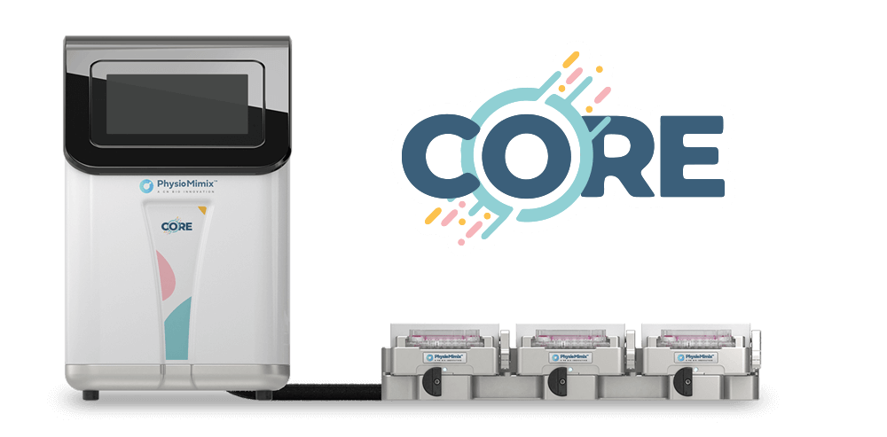
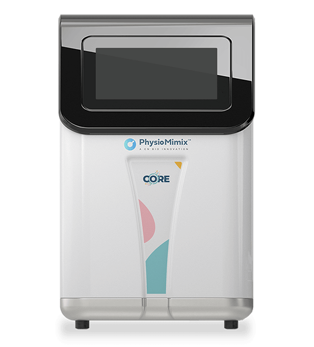
| CN Bio product used | PhysioMimix Core System was used to run the three-dimensional (3D) perfused human nonalcoholic steatohepatitis (NASH) co-culture model. |
| How the platform was used | Primary human hepatocytes were co-cultured with hepatic Kupffer cells and hepatic stellate cells under perfusion in medium enriched in fatty acids, sugars, and insulin to produce a NASH phenotype. Small interfering RNA (siRNA) against SPTBN1 (5 nM, 25 nM, or 50 nM) or control siRNA was added from day 4, every 2 days with medium changes, with transcript and RNA sequencing (RNA-seq) readouts 96 h later at day 8 and lipid staining at day 8 and day 13. |
| Biological context | Human liver; NASH; 3D perfused tri-culture of primary human hepatocytes, hepatic Kupffer cells, and hepatic stellate cells under lipid-loaded and sugar-loaded conditions. The wider paper also covers hepatocyte-specific Sptbn1 knockout mice, diet-induced NASH and hepatocellular carcinoma (HCC) mouse models, and human NASH and HCC liver tissue. |
| Comparator | In the human 3D model: control siRNA (siCtrl) cultures at matched concentrations. Elsewhere in the paper: Flox littermate control mice, siCtrl-injected mice, and healthy obese versus NASH liver expression data from a public dataset (GSE48452). |
| Key readouts | SPTBN1 transcript abundance, RNA-seq with pathway and Ingenuity Pathway Analysis (IPA) upstream regulator analysis, lipid accumulation by Oil Red O normalized to total protein, lactate dehydrogenase (LDH) activity as a cell health marker, and albumin production as a hepatic function marker. Animal and patient arms add serum triglyceride (TG), glucose, aspartate transaminase (AST), alanine transaminase (ALT), liver histology, NAFLD Activity Score (NAS), tumor burden, immunohistochemistry, and single-nucleus RNA-seq. |
| Main interpretation | SPTBN1 supports CASPASE-3-driven SREBP1 activity and pro-fibrotic transforming growth factor β (TGF-β) signaling in the liver, and that silencing it reverses NASH-associated transcriptional programs in mice and in a perfused human 3D liver model, which positions SPTBN1 as a candidate siRNA target for metabolic liver disease. |
Table of Contents
Which CN Bio product was used?
The paper names the PhysioMimix OOC platform (CN Bio Innovations) as the system used for its human 3D NASH culture model. The model is a perfused co-culture of primary human hepatocytes with hepatic Kupffer cells and hepatic stellate cells, the same tri-culture arrangement behind CN Bio’s current Liver-on-a-chip models, maintained in medium enriched in fatty acids, sugars, and insulin for two weeks. The authors cite Kostrzewski et al. (2017, 2020) as the source of the model. Cells were seeded at day 0, moved into the lipid-loaded medium at day 1, transfected with siRNA at day 4 using RNAiMAX at a 1:3 ratio, and analyzed at day 8, with Oil Red O measured at day 8 and day 13. The 3D human NASH co-culture work was performed by a CN Bio author (Gareth Guenigault, CN Bio Innovations, Cambridge).
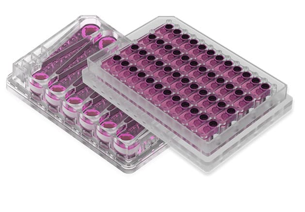
The platform was used for the human in vitro arm of the study only. Mouse work used liver-specific Sptbn1 knockout (LSKO) animals, high-fat diet (HFD) and western diet (WD) feeding, hydrodynamic tail vein delivery of siRNA, and diethylnitrosamine (DEN) induced liver cancer models, while the patient work used immunohistochemistry, single-nucleus RNA-seq, and public expression datasets. Hardware configuration, flow rate, hepatocyte donor number, and donor characteristics are not specified in the paper, and it does not name the consumable format used, so it cannot be mapped onto a particular PhysioMimix Multi-chip Liver plate.
What this paper is about
Nonalcoholic fatty liver disease (NAFLD) and NASH, now termed metabolic dysfunction-associated steatotic liver disease (MASLD) and metabolic dysfunction-associated steatohepatitis (MASH), arise from obesity and metabolic dysfunction, and the paper cites estimates that they affect up to a third of the world’s population. Few single agents reverse both steatosis and fibrosis, so the authors set out to find a molecular node that sits in both processes. Their candidate was SPTBN1 (β2-spectrin), a multidomain adaptor protein already known to promote TGF-β receptor activation of SMAD3 and to be cleaved by CASPASE-3.
The question the study addresses is whether removing SPTBN1 from hepatocytes changes the course of diet-induced liver disease, and by what mechanism. To answer it, the authors generated liver-specific Sptbn1 knockout mice, fed them a high-fat diet or a western diet, ran siRNA knockdown as both a prevention and a treatment experiment, mapped the biochemical interaction between the CASPASE-3 cleavage products of SPTBN1 and SREBP1, and checked the findings against human NASH and HCC tissue. The human 3D liver MPS provided the test of whether silencing SPTBN1 produces the same transcriptional shift in human cells under NASH conditions that it produces in mouse liver, which is the role perfused human models increasingly play in in vitro disease modeling.
What the researchers found
Loss of hepatocyte SPTBN1 protected mice from diet-induced liver disease. LSKO mice on a high-fat diet gained less weight, carried less visceral fat, and showed lower serum TG, AST, and ALT than Flox controls, along with reduced liver TG. Their liver histology showed normal architecture with minimal lipid, no ballooning, and none of the fine collagen deposition seen by Sirius red staining in controls, which produced a lower NAS. Western diet feeding gave the same pattern.
The mechanism runs through CASPASE-3 cleavage of both SPTBN1 and SREBP1. Stress conditions in immortalized human hepatocytes (THLE2) and Huh7 HCC cells activated CASPASE-3, which cleaved SPTBN1 into N-terminal and C-terminal fragments and generated the nuclear form of SREBP1. Only the N-terminal SPTBN1 fragment co-immunoprecipitated with nuclear SREBP1, and molecular docking placed the interaction on SREBP1c residues Q295 to K374, which cover the basic helix-loop-helix domain. Knocking down SPTBN1 reduced nuclear SREBP1 without preventing CASPASE-3 activation, which the authors interpret as SPTBN1 stabilizing the cleaved transcription factor rather than driving its production. SREBP1 target gene expression (Acc1, Scd1, Fasn) fell in LSKO livers, while LDLR, mainly under SREBP2 control in liver, did not change. The effect was independent of SCAP and INSIG, and independent of SMAD3.
siRNA against Sptbn1 worked in mice as both prevention and treatment. Three hydrodynamic injections during high-fat feeding reproduced the LSKO phenotype, with less weight gain, less visceral fat, lower pro-fibrotic and inflammatory gene expression, and no histological signs of NAFLD or NASH. In animals with NASH already established by 12 weeks of western diet, four injections reduced liver weight, NAS, serum TG, glucose, and Mmp2 expression, with steatosis, lobular inflammation, ballooning, and fibrosis all improved on blinded pathology review. Body weight did not change significantly in that treatment arm, which the authors read as a body weight-independent effect.
Human NASH tissue matched the mouse mechanism. Immunohistochemistry showed increased cleaved CASPASE-3 in NASH hepatocytes alongside significantly increased nuclear SPTBN1 (p < 0.05) and increased nuclear SREBP1, both of which are mainly cytoplasmic in healthy controls. In a public dataset comparing 16 healthy obese individuals with 17 NASH patients, SPTBN1 and CASPASE-3 transcripts were higher in NASH while SREBP1 itself was unchanged, and the SREBP1 targets FASN, SCD1, and AACS were higher, which the authors read as increased SREBP1 activity rather than increased SREBP1 expression.
In the PhysioMimix 3D human NASH model, siRNA against SPTBN1 reversed the NASH transcriptional signature. Treatment at 25 nM reduced SPTBN1 transcripts by more than 90% at 96 h with no significant change in LDH activity or albumin production, so the knockdown was achieved without loss of cell health or hepatic function. Oil Red O staining showed a small decrease in lipid accumulation at day 8 that did not reach significance. RNA-seq at 25 nM and 50 nM showed lower expression of genes covering lipid transport, triglyceride and glycogen metabolism, and lipoprotein catabolism, together with SREBP1 target genes, plus reduced fibrosis-associated transcripts, altered inflammatory gene expression, and a decreased TGF-β signaling signature. IPA upstream regulator analysis produced the clearest result: regulators with higher activity in human NASH than in NAFLD had lower activity in siSPTBN1-treated cultures, and regulators with lower activity in NASH had higher activity after treatment. The tri-culture used here is now packaged for laboratories that want to run it in house as NASH-in-a-box.
LSKO mice developed fewer liver tumors. Combined DEN and western diet feeding produced liver tumors in Flox mice but not in LSKO mice, with lower liver weight, fewer visible nodules, and less Ki67 labeling. With DEN alone, all mice developed tumors, but LSKO animals had fewer and smaller ones. In TCGA data, homozygous SPTBN1 deletion was rare (1 of 440 HCC cases, and 0.1% across 30 cancer types), while higher SREBP1 target gene expression in HCC was associated with worse overall and disease-free survival (p < 0.05).
What the researchers found
The main finding was that modest, physiologically relevant hyperinsulinemia was sufficient to induce key features of hepatic insulin resistance in perfused primary human hepatocytes. Hepatocytes exposed to 800 pM insulin showed reduced insulin clearance over time, impaired AKT phosphorylation, and reduced insulin-mediated suppression of hepatic glucose production.
The study showed that elevated glucose and free fatty acids compounded the insulin-resistant phenotype. The combined disease-like condition, referred to as Condition 2, produced the strongest metabolic impairment, with increased hepatic glucose production, impaired repression of PCK1 and G6PC, and reduced insulin uptake driven primarily by hyperinsulinemia.
Condition 2 also reproduced several early MASLD-associated phenotypes. The model showed increased intracellular triglycerides, elevated taurine- and glycine-conjugated cholic acid species, and secretion of inflammatory chemokines including CCL2, IL8, CXCL1, and CXCL10, without significant increases in alanine aminotransferase, aspartate aminotransferase, or lactate dehydrogenase.
Transcriptomic analysis identified 914 differentially expressed genes between the physiological baseline and Condition 2. The disease-like condition aligned with human liver and metabolic disease signatures, including acquired metabolic disease, cholestasis, type 2 diabetes, bile acid synthesis and transport, and inflammatory signaling.
The intervention experiments showed partial reversibility. Returning disease-like hepatocytes to baseline media normalized hepatic glucose production and insulin uptake, but did not reverse intracellular triglyceride accumulation. Resmetirom improved insulin sensitivity, normalized steatosis in male and female hepatocytes, and activated the target gene DIO1.
The paradoxical result was that resmetirom increased CXCL1 and IL8 secretion in both baseline and disease-like hepatocytes, with a stronger effect in Condition 2. Label-free imaging and gene expression data suggested that increased mitochondrial activity or stress, reflected by higher nicotinamide adenine dinucleotide phosphate signal and increased PGC-1α and SOD2 expression, may contribute to this inflammatory response.
Why the paper matters
Target validation for liver disease usually stalls at the point where a mouse phenotype has to be shown to hold in human cells. This paper handles that step with a perfused human liver MPS rather than a monolayer assay, and the readout is a whole transcriptional program rather than a single biomarker. That matters for anyone deciding what evidence a target needs before it moves forward: the IPA upstream regulator comparison shows the human model responding to knockdown in the direction opposite to human NASH progression, which is a stronger form of evidence than a single reduction in lipid content.
The paper is also useful as a worked example of testing an oligonucleotide in a human 3D liver model. Knockdown above 90% was achieved with repeated dosing into a perfused culture while LDH activity and albumin production held steady, so the transcriptional changes can be read as target engagement rather than toxicity. The lipid staining result is a fair counterweight: a four-day treatment window in a two-week culture moved gene expression clearly but moved measured lipid only slightly, which is a practical point about endpoint selection and study duration. Later versions of the assay pair the transcriptional picture with secreted fibrosis, inflammation, and steatosis biomarkers over at least 14 days, as described on the PhysioMimix MASH assay page. For groups building a case to reduce animal use, the paper shows a human MPS carrying the confirmatory role for a target defined in mice, in a disease where mouse models are known to translate poorly.
Find out more about how perfusion and 3D scaffold culture support this kind of study on the organ-on-a-chip technology page.
Key study takeaways
- The study used the PhysioMimix OOC platform to run a perfused 3D co-culture of primary human hepatocytes, hepatic Kupffer cells, and hepatic stellate cells as a human NASH model.
- The model reproduced a NASH-associated transcriptional state that could be reversed: siRNA against SPTBN1 lowered fatty acid metabolism genes, SREBP1 target genes, fibrosis genes, and a TGF-β signaling signature.
- Compared with control siRNA cultures, siSPTBN1 cultures showed more than 90% SPTBN1 knockdown at 25 nM with no significant change in LDH activity or albumin production, and a non-significant reduction in Oil Red O staining.
- The workflow combined transcript quantification, RNA-seq with IPA upstream regulator analysis, Oil Red O lipid staining, LDH activity, and albumin measurement from the same perfused cultures.
- The findings support the use of perfused human liver MPS models for confirming targets and mechanisms identified in rodent models of metabolic liver disease, and for testing siRNA in a human cell context.
Why this paper is worth reading
This paper is useful because it shows what a perfused human liver model contributes at a specific decision point: whether a target validated in knockout mice behaves the same way in human liver cells held under disease conditions. Researchers working on metabolic liver disease targets, oligonucleotide therapeutics, or chronic liver disease models will find a clear template for pairing rodent genetics with a human organ-on-a-chip readout, along with an honest account of which endpoints moved and which did not. Read it alongside the model characterization papers by Kostrzewski et al. that it cites, which set out how the tri-culture behaves before a therapeutic is added.
FAQ
The study used the PhysioMimix OOC platform from CN Bio Innovations to run its 3D human NASH co-culture model. The specific PhysioMimix hardware configuration and multi-chip plate type are not specified in the paper.
The PhysioMimix platform maintained a perfused 3D co-culture of primary human hepatocytes, hepatic Kupffer cells, and hepatic stellate cells in medium enriched in fatty acids, sugars, and insulin to generate a NASH phenotype. siRNA targeting SPTBN1 at 5 nM, 25 nM, or 50 nM was added from day 4 every 2 days with medium changes, with analysis at day 8 and Oil Red O staining at day 8 and day 13. Flow rate is not specified.
What biological model or disease area was studied?
The disease areas are nonalcoholic steatohepatitis (NASH) and hepatocellular carcinoma (HCC). The human model was a perfused 3D liver microphysiological system built from primary human hepatocytes with hepatic Kupffer cells and hepatic stellate cells. The paper also used liver-specific Sptbn1 knockout mice, diet-induced and DEN-induced mouse models, and human NASH and HCC liver tissue.
SPTBN1 promotes CASPASE-3-dependent SREBP1 activity and de novo lipogenesis in the liver, and removing or silencing SPTBN1 protected mice from diet-induced obesity, NASH, and liver tumors. In the PhysioMimix 3D human NASH model, siRNA against SPTBN1 reversed NASH-associated transcriptional changes in fatty acid metabolism, fibrosis, and TGF-β signaling.
In the PhysioMimix model, siSPTBN1-treated cultures were compared with control siRNA cultures at matched concentrations. In the animal work, LSKO mice were compared with Flox littermate controls and siSptbn1-treated mice with siCtrl-treated mice. In the human tissue work, NASH samples were compared with healthy control and healthy obese samples.
The PhysioMimix arm used SPTBN1 transcript abundance, RNA-seq with pathway and IPA upstream regulator analysis, Oil Red O staining normalized to total protein, LDH activity, and albumin production. The wider study added serum TG, glucose and cholesterol, AST and ALT, liver TG, histology and NAS scoring, immunoblotting, immunohistochemistry, proximity ligation assays, luciferase reporter assays, single-nucleus RNA-seq, and tumor burden.
The paper shows a perfused human liver MPS being used to confirm, in human cells, a target identified through mouse genetics, and it reports both the endpoints that responded (transcriptional programs for lipogenesis, fibrosis, and TGF-β signaling) and the endpoint that did not reach significance (lipid accumulation by Oil Red O at day 8). That makes it a practical reference for target validation strategy, siRNA testing in 3D human liver models, and endpoint and duration choices in NASH and MASH model design.
Full citation
Related products and services
Contract research services
Discover how to utilize our cross-species models to inform next-step decision making via our DILI in vitro Contract Research Services here.
Add PhysioMimix Core in your lab
To develop your own cross-species Liver MPS models, you will need: