

Resource > Scientific publications >
Protocol for establishing rectal tumor organoid-derived monolayers in a microphysiological system to model radiotherapy
Filed under: Disease modeling and Oncology
Summary
Researchers at the Belgian Nuclear Research Center (SCK CEN) and collaborating universities used the PhysioMimix Barrier plate with the PhysioMimix Core System to perfuse rectal PDTO-derived monolayers on Transwell inserts while delivering five daily fractions of X-ray irradiation, reproducing the fractionated scheduling used in rectal cancer radiotherapy. Monolayers reached confluence at around 10 days, and TEER increased to a plateau and then settled between 1,500 and 2,500 Ω·cm² as the monolayer differentiated. Whole-proteome analysis identified and quantified 1,726 proteins with irradiation-linked clustering into programmed cell death, cellular stress response, and mitotic cell cycle regulation pathways. For groups building radiotherapy or radiotoxicity models, this protocol provides a documented human in vitro workflow covering organoid subculture, monolayer seeding, perfusion, and fractionated irradiation, as an alternative to mouse models and immortalized cell lines, which the authors say do not adequately represent human-specific epithelial damage and radiotoxicity responses.
Study facts at a glance
| Publication | Bouges E, Segers C, Lezzerini M, Leys N, Lebeer S, Zhang J, Mastroleo F. Protocol for establishing rectal tumor organoid-derived monolayers in a microphysiological system to model radiotherapy. STAR Protocols. September 18, 2026;7:104708. |
| DOI | 10.1016/j.xpro.2026.104708 |
| CN Bio product used | PhysioMimix Barrier plate and the PhysioMimix Core System |
| How the platform was used | Transwell® inserts carrying rectal patient-derived tumor organoid (PDTO) monolayers were transferred into the PhysioMimix Barrier plate on day 14, after the plate was primed with 750 µL of core medium per well at 1 µL/s for at least 24 h. Culture then ran at 0.5 µL/s at 37°C and 5% CO2 for five consecutive days of daily X-ray irradiation, with flow paused for each transepithelial electrical resistance (TEER) reading, medium change, and irradiation run. |
| Biological context | Human rectal adenocarcinoma. The commercially available PDTO line HCM-CSHL-0143-C20 (PDM-96™, ATCC), from an 88-year-old female donor with stage 1 colorectal cancer, was expanded in Matrigel® and seeded as 2D monolayers on Matrigel-coated 0.4 µm Transwell inserts, on either the apical or the basolateral face. Tumor tissue only, with no matched healthy tissue. |
| Comparator | Static Transwell monolayers for the histology comparison, unirradiated 0 Gy monolayers for the proteomics comparison, three seeding densities for barrier formation, and published Caco-2 and HT-29 monolayer TEER values as a literature benchmark. |
| Key readouts | TEER, brightfield imaging of confluence, hematoxylin and eosin staining of cryosections for thickness and stratification, whole-proteome analysis with Reactome pathway clustering, protein quantification by bicinchoninic acid (BCA) assay, and RNA yield with RNA integrity number (RIN) scoring. |
| Main interpretation | Rectal PDTO-derived monolayers can be held under perfusion in the PhysioMimix Barrier plate and given a fractionated irradiation course rather than a single dose, producing barrier, histology, and proteomic readouts from human tumor tissue within a working window of roughly five to seven days at full confluence. |
Table of Contents
Which CN Bio product was used?
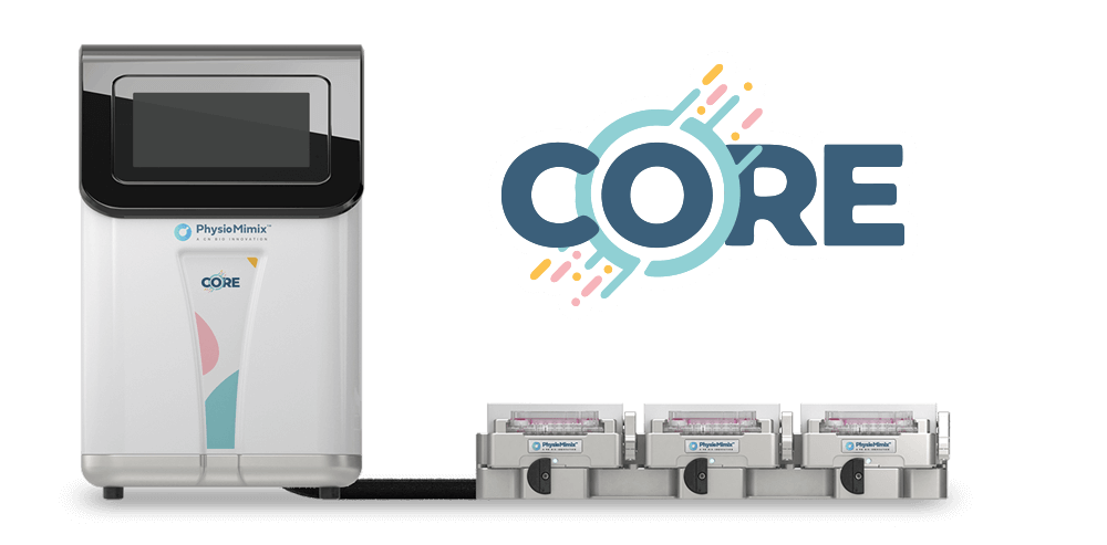
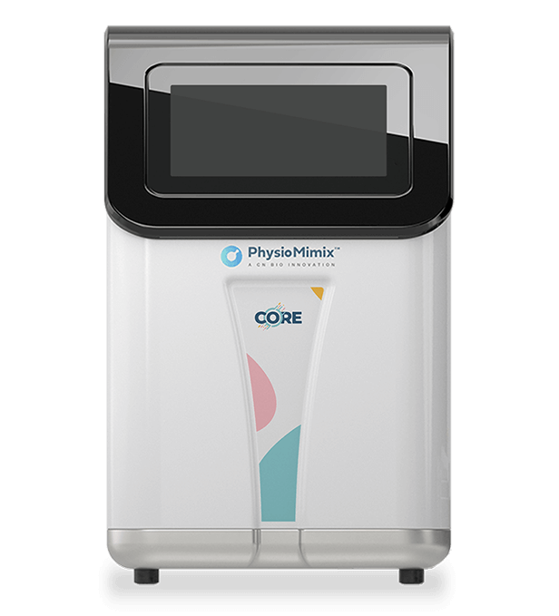
The study used the PhysioMimix Barrier plate together with the PhysioMimix Core System. On day 13 the sterile barrier plate was seated in the driver and primed with 750 µL of pre-warmed core medium per well, then run on the incubate setting at 1 µL/s for a minimum of 24 h to wet the channels and clear air bubbles. On day 14 the priming medium was replaced with 750 µL of fresh core medium per well, the Transwell inserts holding the mature monolayers were moved into the plate wells with forceps, 250 µL of core medium was added to the apical side of each insert, and the driver returned to the docking station inside the incubator at a flow rate of 0.5 µL/s, selected by the authors to give biologically relevant fluid dynamics. Flow was paused before every TEER measurement, medium change, and irradiation run, and the plate stayed in the driver until the end of the experiment.
The PhysioMimix platform covers the perfusion phase only, days 13 to 18 of the monolayer workflow. Organoid subculture, monolayer seeding on Transwell inserts, and the roughly two weeks of monolayer maturation were carried out in standard 12-well and 24-well plates, and irradiation was delivered off-platform using an Xstrahl 320 kV tube (dose rate 8 Gy/h, tube voltage 250 kV, tube current 12 mA, vertical beam orientation.
What this paper is about
Colorectal cancer is described by the authors as the third most prevalent cancer worldwide, and radiotherapy forms part of standard clinical protocols for rectal disease. The gap they identify is a modeling one: conventional preclinical models of the gastrointestinal tract, meaning mice and 2D immortalized cell lines, do not adequately represent human-specific epithelial damage and radiotoxicity responses. Existing in vitro radiobiology work typically relies on immortalized lines or static organoid cultures given a single radiation dose, which does not reflect the fractionated schedules used clinically.
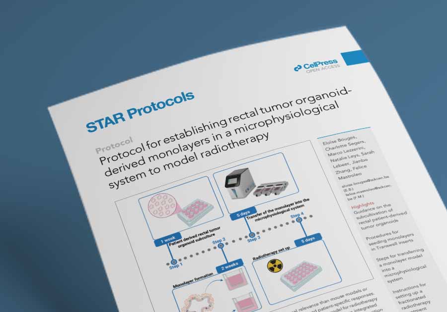
The protocol answers that with a human rectal tumor model built in four stages: subculture of patient-derived tumor organoids in Matrigel drops, dissociation of the organoids and seeding of the fragments as monolayers on Matrigel-coated Transwell inserts, transfer of the mature inserts into the PhysioMimix Barrier plate, and five consecutive daily X-ray fractions. PDTOs were chosen because they preserve the cellular heterogeneity of the source tissue and, as earlier work on rectal cancer organoids showed, can reflect patient-specific response to chemoradiation. Monolayers were chosen because they give experimental access to both the apical and the basolateral face of the epithelium.
Two seeding configurations are described side by side. Monolayers seeded on the basolateral face of the Transwell insert sit directly in the perfused well, so flow passes their apical surface, modeling the lumen side of the rectal epithelium. Monolayers seeded on the apical face experience flow on their basolateral surface, modeling conditions at the lamina propria. Both were compared against static Transwell controls, and irradiated monolayers were compared against unirradiated 0 Gy controls. The applications named by the authors are radiotherapy modeling, toxicity testing, and adjuvant therapy screening.
What the researchers found
The study reported that fractionated irradiation at 4 and 6 Gy produced significant proteomic changes. Whole-proteome analysis of the monolayers identified and quantified 1,726 proteins, and functional clustering using Reactome grouped the response into pathways related to programmed cell death, cellular stress response, and mitotic cell cycle regulation. Upregulated proteins were primarily associated with stress adaptation and apoptosis, while downregulated proteins were linked to cell cycle progression. The protocol involved the use of 2 Gy fractions for a cumulative irradiation dose of 10 G, and the reported experiments covered 0, 4, 6, and 8 Gy dosing.
Barrier formation followed a reproducible pattern. Brightfield imaging showed confluence typically within approximately 10 days of seeding, with the objective of reaching full confluence before irradiation begins on day 14. TEER values rose progressively to a plateau indicating full confluence, then decreased, which the authors attribute to monolayer differentiation and tight junction maturation, settling between 1,500 and 2,500 Ω·cm². The authors note that this sits below the values above 2,500 Ω·cm² reported for Caco-2 and HT-29 derived monolayers, and read the PDTO profile as giving further physiological relevance to the model.
Seeding density set the timing. At 2×10⁴ cells per 0.33 cm² insert, full confluence arrived at around day 10 as TEER increased gradually to a plateau. At 8.84×10⁴ cells per 0.33 cm², confluence arrived at around day 6, with TEER reaching an initial plateau, then falling and stabilizing at a lower secondary plateau, likely reflecting monolayer differentiation. At the recommended 3×10⁴ cells per insert, the plateau was reached at day 12 and values decreased after day 15.
Histology separated the two flow configurations. Five days of apical flow induced a trend of increased monolayer thickness compared with static model monolayers, while flow applied to the basolateral side had minimal effect on thickness. Stratification was more pronounced in both apical and basolateral MPS cultures than in their respective static controls, which was taken as confirmation that dynamic conditions can promote tissue-like architecture.
The protocol also quantified how much material each insert format returns. One 1.12 cm² Transwell insert served as a single biological replicate for proteomics, while three pooled 0.33 cm² inserts were required to obtain sufficient starting material. For transcriptomics, three pooled 0.33 cm² inserts delivered 8.4±2.4 µg of RNA with RIN values above 9, leading the to the conclusion that even one 0.33 cm² insert may be sufficient for RNA sequencing. Recovery of viable organoids depended on handling detail that the troubleshooting section makes explicit. Swing-bucket centrifugation, pipette tips coated with 0.1% OncoPro BSA, and Protein LoBind tubes were all needed to avoid significant material loss at thawing and splitting, and cultures run without them showed poor survival and limited expansion. Even distribution of fragments across the insert at the time of seeding predicted whether monolayers reached confluence. Basolateral seeding lost fragments to gravity when the inserts were inverted back into the wells, and the authors compensated by seeding at 4×10⁵ cells per mL instead of 3×10⁵ cells per mL.
Why the paper matters
For groups planning radiotherapy or radiotoxicity work, the practical value sits in the handling detail. Rectal PDTOs are lost easily at thawing and splitting, and the protocol names the specific causes and the fixes rather than leaving them to be rediscovered. It also supplies the numbers needed before committing donor material: cells per insert, days to confluence, expected TEER range, protein and RNA yield by insert format, and how many inserts to pool for each analysis.
The dosing schedule is the second point. A single dose delivered to a static culture is a poor stand-in for a clinical course of fractions, and running five daily fractions in a perfused barrier plate keeps the model measurable across the whole course, with TEER recorded before each fraction and spent media available for further analysis such as an LDH assay. The paper is also direct about where the model stops. Rectal epithelium renews quickly, so a fully confluent monolayer cannot be maintained for more than 5 to 7 days without affecting human relevance, which caps the length of any treatment response study run this way. The model lacks key features of the gut microenvironment, including the characteristic oxygen gradient and immune system components, and the authors flag radioprotective biotic agents and immune cells as work still to be done. Inter-batch and patient-to-patient variability can occur, particularly in TEER values and time to confluence. The supporting dataset is part of a broader study still in progress.
Key study takeaways
- The study used the PhysioMimix Barrier plate with a PhysioMimix Core System to perfuse rectal PDTO-derived monolayers on Transwell inserts at 0.5 µL/s across five consecutive daily X-ray fractions.
- The monolayers formed a measurable epithelial barrier, reaching confluence at around 10 days and settling at TEER values between 1,500 and 2,500 Ω·cm² after differentiation.
- Compared with static Transwell monolayers, monolayers under apical flow showed a trend of increased thickness, and stratification was more pronounced under both apical and basolateral flow.
- The workflow combined TEER, brightfield imaging, hematoxylin and eosin histology of cryosections, whole-proteome analysis, and RNA yield and integrity checks.
- Fractionated irradiation at 4 and 6 Gy produced significant proteomic changes across 1,726 quantified proteins, supporting the use of PDTO-derived barrier models for radiotherapy response and radiotoxicity studies.
- The paper indicates the model is most appropriate for studies that fit inside a 5 to 7 day window at full confluence, and that it carries no oxygen gradient or immune component.
Why this paper is worth reading
This paper is useful because it publishes the operational detail behind a fractionated radiotherapy experiment: volumes, flow rates, seeding densities, day-by-day timings, and five documented troubleshooting problems that decide whether a rectal PDTO monolayer survives long enough to irradiate. A scientist deciding whether to build a fractionated radiotherapy model in a perfused barrier plate can use it to estimate how many inserts to run, when confluence will arrive, what TEER to expect, and what proteomic or transcriptomic material will come out at the end. The apical and basolateral seeding comparison deserves attention on its own, since the two configurations place flow on opposite faces of the epithelium and behave differently in both barrier maturation and histology.
FAQs
The study used the PhysioMimix Barrier plate and the PhysioMimix Core System.
The PhysioMimix Barrier plate was primed with core medium at 1 µL/s for at least 24 h, then loaded with Transwell inserts carrying rectal PDTO-derived monolayers and perfused at 0.5 µL/s for 5 days of daily fractionated X-ray irradiation. Flow through the PhysioMimix Barrier plate was paused for each TEER reading, medium change, and irradiation run.
The disease area is human rectal adenocarcinoma. Patient-derived tumor organoids from the ATCC line HCM-CSHL-0143-C20 (PDM-96), taken from an 88-year-old female donor with stage 1 colorectal cancer, were dissociated and seeded as 2D monolayers on Matrigel-coated Transwell inserts, then perfused in the PhysioMimix Barrier plate.
Rectal PDTO-derived monolayers can be grown to a stable epithelial barrier, transferred into the PhysioMimix Barrier plate, and exposed to five daily radiation fractions while remaining measurable by TEER, histology, and proteomics. Fractionated irradiation at 4 and 6 Gy produced significant proteomic changes across 1,726 quantified proteins, clustering into programmed cell death, cellular stress response, and mitotic cell cycle regulation pathways.
The study compared perfused monolayers in the PhysioMimix Barrier plate against static Transwell monolayers by histology, irradiated monolayers against unirradiated 0 Gy controls by proteomics, apical seeding against basolateral seeding, and three seeding densities by TEER. Published Caco-2 and HT-29 monolayer TEER values were used as a literature benchmark.
The readouts were TEER, brightfield imaging of confluence, hematoxylin and eosin staining of cryosections for thickness and stratification, whole-proteome analysis with Reactome pathway clustering, protein quantification by BCA assay, and RNA yield with RIN scoring.
The paper gives researchers a documented human in vitro route to fractionated radiotherapy modeling in rectal tumor tissue, with the seeding densities, flow rates, timings, expected TEER values, and per-insert material yields needed to plan an experiment. It also states where the rectal PDTO model stops being appropriate: full confluence holds for only five to seven days, and the model has no oxygen gradient or immune component.
Full citation
Related products and services
Contract research services
Discover how to utilize our cross-species models to inform next-step decision making via our DILI in vitro Contract Research Services here.
Add PhysioMimix Core in your lab
To develop your own cross-species Liver MPS models, you will need: