

Resource > Scientific publications >
Gut-Liver Physiomimetics Reveal Paradoxical Modulation of IBD-Related Inflammation by Short-Chain Fatty Acids
Filed under: Disease modeling and Immune-mediated liver injury
Summary
Trapecar and colleagues at MIT combined a perfused primary human liver MPS, cultured on the same 301-channel scaffold format used in CN Bio’s Legacy LiverChip®, with a colon MPS built from ulcerative colitis (UC) patient organoids and with circulating regulatory and effector CD4 T cells, then tested how microbiome-derived short-chain fatty acids (SCFAs) act across the human gut-liver axis. Fluidic coupling of the two organs on its own increased hepatic metabolism and lowered inflammatory signaling in both tissues, and apically dosed SCFAs further reduced innate immune activation in the UC gut while increasing bile acid synthesis, gluconeogenesis, and ketone body production in the liver. Adding activated Treg and Th17 cells reversed that picture at the same SCFA dose, producing complete gut barrier failure, stalled hepatic albumin production, and higher effector cytokine release, which offers a mechanistic reading of why SCFA supplementation trials in IBD have returned conflicting results.
Which CN Bio product was used?
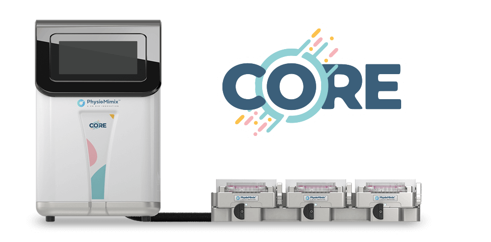
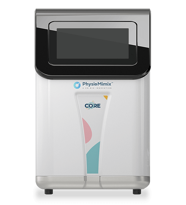
They used a Legacy LiverChip (replaced by PhysioMimix® LC12 and PhysioMimix Core System) for liver MPS experiments run in isolation. Liver tissue in both settings was formed the same way: primary human hepatocytes and Kupffer cells at a 10:1 ratio on collagen I-coated polystyrene scaffolds, 0.25 mm thick and perforated with 301 channels of 0.3 mm diameter, seeded three days before the interaction studies began and maintained under continuous perfusion. Hepatic function was tracked by albumin secretion measured at every 48-hour medium change.
Gut-liver and gut-liver-immune interaction studies used 3XGL, a platform designed at MIT.
Study facts at a glance
| Publication | Trapecar M, Communal C, Velazquez J, Maass CA, Huang YJ, Schneider K, Wright CW, Butty V, Eng G, Yilmaz O, Trumper D, Griffith LG. Gut-Liver Physiomimetics Reveal Paradoxical Modulation of IBD-Related Inflammation by Short-Chain Fatty Acids. Cell Systems. 2020 Mar 25;10:223-239. |
| DOI | 10.1016/j.cels.2020.02.008 |
| CN Bio product used | Legacy LiverChip® (CN Bio Innovations), used to culture the MPS in isolation. |
| How the platform was used | Primary human hepatocytes and Kupffer cells were seeded at a 10:1 ratio (600,000 and 60,000 cells per well) onto collagen I-coated polystyrene scaffolds perforated with 301 channels, then perfused either in the LiverChip for liver-only experiments or in the 3XGL platform for 4-day gut-liver interaction studies with recirculating flow between compartments. |
| Biological context | Human gut-liver axis in ulcerative colitis (UC), a form of inflammatory bowel disease (IBD). The gut MPS used colon organoid-derived epithelial monolayers from a UC donor and a non-diseased control donor on Transwell® inserts, with monocyte-derived macrophages and dendritic cells attached to the basolateral surface. The liver MPS used primary human hepatocytes with Kupffer cells. CD4 regulatory T cells (Tregs) and T helper 17 (Th17) cells were added to circulation at a 2:1 ratio. All cells were human and donor-mismatched. |
| Comparator | Static off-platform Transwell culture, UC versus non-diseased epithelium, gut MPS in isolation versus fluidically coupled to the liver MPS, with and without 20 mM total short-chain fatty acids (SCFAs), and with and without circulating Treg and Th17 cells. |
| Key readouts | Insulin uptake, hepatic glucose production, AKT RNA sequencing with GEO, Gene Ontology (GO), KEGG, and REACTOME pathway enrichment; transepithelial electrical resistance (TEER); 41-plex cytokine and chemokine panels plus transforming growth factor beta (TGF-β) and Th17 panels; targeted SCFA quantification and global metabolomics by liquid chromatography-tandem mass spectrometry; hepatic albumin secretion; confocal immunofluorescence; physiologically based pharmacokinetic (PBPK) modeling; Random Forest classification. |
| Main interpretation | The direction of SCFA activity on inflamed colon tissue depended on the activation state of CD4 T cells: SCFAs reduced innate immune activation in the UC gut MPS and increased hepatic metabolic function, but worsened barrier failure and liver injury once effector T cells were engaged. |
Table of Contents
Which CN Bio product was used?
The LiverChip was used as the perfused culture system used for liver MPS experiments run in isolation. Liver tissue in both settings was formed the same way: primary human hepatocytes and Kupffer cells at a 10:1 ratio on collagen I-coated polystyrene scaffolds, 0.25 mm thick and perforated with 301 channels of 0.3 mm diameter, seeded three days before the interaction studies began and maintained under continuous perfusion. Hepatic function was tracked by albumin secretion measured at every 48-h medium change.
The multi-organ gut-liver and gut-liver-immune interaction studies used 3XGL, a platform designed at MIT. The LiverChip is the forerunner of the PhysioMimix® Core Organ-on-a-chip System, so this study sits in the technical lineage of the current Liver-on-a-chip and Gut/Liver-on-a-chip models rather than being a study performed on them.
Find out more about CN Bio Liver-on-a-chip models here
Find out more about CN Bio DILI assays here
What this paper is about
Clinical observation ties inflammatory bowel disease to inflammatory liver disease, with up to 80% of patients who have primary sclerosing cholangitis (PSC) also having concurrent UC, yet the direction of cause and effect has stayed unresolved. Animal models give poor control over experimental variables, and conventional in vitro models leave out the organ-organ traffic that shapes how gut-derived molecules reach and act on the liver. SCFAs sit at the center of the disagreement. Acetate, propionate, and butyrate, produced by microbial fermentation of fiber in the colon, have been credited with strengthening the gut barrier and promoting immune tolerance, while other work reports that they push CD4 and CD8 T cells toward effector function, and several human and animal trials have found no benefit or outright worsening of inflammation.
The authors built an all-human, three-part model to separate those effects. A gut MPS of ulcerative colitis (primary UC colon epithelium with monocyte-derived macrophages and dendritic cells) was connected to a liver MPS (primary hepatocytes with Kupffer cells) under recirculating flow, with Treg and Th17 cells circulating between compartments. Physical contact between circulating T cells and the epithelium was blocked by the microporous Transwell membrane, while the liver scaffold allowed T cells to enter the tissue directly, which mirrors hepatic trafficking of peripheral T cells. Three modes of operation were compared: gut MPS alone, gut plus liver, and gut plus liver plus circulating T cells, each with and without 20 mM total SCFAs dosed apically at a physiological 6:2:2 molar ratio.
What the researchers found
The UC and control gut MPSs diverged before any SCFA was added. Compared with the non-diseased donor, UC epithelia showed disease-associated pathway enrichment against GEO, upregulated innate immune activation, apoptotic signaling, and epidermal growth factor activity by GO, and increased MAPK and PI3K-Akt signaling by KEGG. Both donors formed confluent monolayers with comparable TEER, but only the UC monolayers showed enlarged hyperchromatic nuclei and irregular actin fragmentation, which are recognized indicators of IBD dysplasia. Co-culture with Treg and Th17 cells at a 2:1 ratio pushed CD4 T cells toward Th1, Th2, and Th17 cytokine production in the UC condition, with increased IL-4, IL-5, IL-13, tumor necrosis factor alpha (TNF-α), and interferons, while the Treg and Th17 balance held with the healthy donor.
SCFA handling by the UC gut MPS tracked human clinical data closely. Over 48 h the epithelium consumed 54% of the applied butyrate, while 96% of acetate and 82% of propionate crossed into the basal compartment, shifting the molar ratio to roughly 7:2:1 in the receiving compartment. That mirrors the reported behavior of butyrate as the primary energy source of colonocytes, of which about half reaches the portal vein. Time-course RNA sequencing at 4, 8, 24, and 48 h showed downregulation of cell cycle and WNT signaling through FoxO, upregulation of hormone receptor activity and peroxisome proliferator-activated receptor (PPAR) signaling, and suppression of nuclear factor kappa B (NF-κB) and major histocompatibility complex class II signaling.
Coupling the two organs changed both of them, before SCFAs entered the picture. The gut MPS increased pentose and glucuronate interconversions, ABC transporters, and protein digestion and absorption, and reduced colorectal cancer-related signaling. The liver MPS increased cytochrome P450 xenobiotic metabolism, glutathione metabolism, biosynthesis of unsaturated fatty acids, and steroid hormone biosynthesis, with downregulation of complement and coagulation cascades, chemokine signaling, and antigen processing and presentation.
Adding SCFAs to the coupled system produced a coherent anti-inflammatory and pro-metabolic result. Gut tissue kept its PPAR upregulation and NF-κB suppression, TEER stayed inside the 100 to 400 Ω/cm² range considered physiological, and hepatic albumin production rose. Liver tissue increased gluconeogenesis, retinol and tyrosine metabolism, and primary bile acid biosynthesis and secretion. Inflammatory mediators in the shared circulating medium fell significantly, with platelet-derived growth factor, IL-9, GRO, and macrophage-derived chemokine emerging as the most condition-predictive reductions by Random Forest. Global metabolomics of the common medium showed enrichment of fatty acid metabolism, ketone body synthesis, and creatine metabolism.
Introducing circulating Treg and Th17 cells converted the UC model into an acute inflammatory one. T cell activation driven by the UC gut MPS reduced gut barrier function and hepatic albumin secretion, upregulated interferon-induced genes in both tissues, and enriched Th1 and Th2 differentiation, IBD, and NOD signaling pathways in the gut alongside Th17 differentiation, allograft rejection, and IBD pathways in the liver. Gene clustering indicated partial engraftment of T cells within liver tissue based on FOXP3, ICOS, and CD28 expression. IFN-γ, TNF-α, IL-1α, and RANTES all increased, and sphingolipid enrichment appeared, matching serum markers reported for autoimmune hepatitis.
Against expectation, adding the same 20 mM SCFA dose to that inflamed condition made everything worse. NF-κB, TNF, and mTOR signaling were strongly enriched in the gut MPS, TEER collapsed to complete barrier failure rather than the partial loss seen with T cells alone, and major hepatic metabolic pathways were broadly downregulated. IFN-γ, IL-5, IL-8, IL-10, and IL-13 were the most condition-predictive cytokines, and measured IFN-γ and TNF-α concentrations sat close to levels reported in serum from patients with IBD. A separate static experiment isolated the mechanism: at 1 mM total SCFA, unstimulated Treg and Th17 cells increased TGF-β1 production, consistent with a tolerance-promoting role, whereas cells stimulated with IL-12 and IFN-γ responded to the same SCFAs with higher IFN-γ, TNF-α, and particularly IL-13 output, alongside REACTOME-level downregulation of histone deacetylase activity and p62-TRAF6 autophagy pathways found only in the activated, SCFA-treated group.
Why the paper matters
Anyone choosing a model for gut-liver or microbiome-immune questions gets a usable decision framework from the methods section. The authors set out why the standard Caco-2 and HT-29 lines are unsuitable for SCFA work, since butyrate inhibits colonic tumor cells and would confound the readout, and why an hTERT-transformed colonocyte alternative did not fit either, because its culture medium is incompatible with primary liver cells and likely with immune cells given the corticosteroid concentrations involved. Their answer was primary patient-derived colon epithelium, accepting a 4 to 8 day working lifespan that matches in vivo intestinal turnover. Anyone reproducing this design inherits both the constraint and the reasoning behind it.
The result also argues for multi-organ configurations over single-organ ones on evidential grounds rather than on principle. Every measurement taken in the gut MPS alone pointed toward SCFAs being anti-inflammatory. The opposite conclusion only became visible once a liver compartment and a circulating adaptive immune compartment were present, and it depended on the T cells being activated. A model without those elements would have returned a confident and wrong answer. That is the practical case for the multi-organ approach in disease modeling work where immune and metabolic axes interact.
There is a reduction argument in here too. Acute T cell-mediated gut and liver inflammation arose from the disease-donor tissue itself, without xenopeptides or the chemical inducers routinely used in animal models of IBD and autoimmune hepatitis.
Key study takeaways
- The liver MPS in this study was cultured on the perfused 301-channel scaffold format used on the Legacy LiverChip, with primary human hepatocytes and Kupffer cells at a 10:1 ratio; the gut-liver interaction studies ran on the MIT-built 3XGL platform.
- The UC gut MPS reproduced disease-relevant behavior, including IBD-associated pathway enrichment, dysplasia-like nuclear and actin morphology, and induction of Th1, Th2, and Th17 cytokine production in co-cultured CD4 T cells that did not occur with healthy donor epithelium.
- SCFA transport across the model matched human data: 54% of butyrate was consumed by the colonic epithelium while most acetate and propionate crossed the barrier, shifting the molar ratio toward the 7:2:1 pattern reported for portal blood.
- Compared with the gut MPS in isolation, fluidic coupling to the liver MPS increased cytochrome P450 and lipid metabolism in the liver and lowered inflammatory pathway expression in both tissues.
- The workflow combined RNA sequencing with pathway enrichment, TEER, multiplex cytokine and chemokine panels, targeted and global metabolomics, albumin secretion, and PBPK modeling of SCFA distribution and clearance.
- SCFAs reduced innate inflammation in the UC gut MPS but worsened barrier failure and hepatic injury once effector CD4 T cells were activated, so the model is most informative when the immune activation state is defined and controlled as an experimental variable.
Why this paper is worth reading
This paper is useful because it shows a multi-organ MPS producing a result that contradicts the single-organ result, and then tracing the contradiction to a specific and testable mechanism in circulating T cells. For anyone weighing whether the added cost and complexity of a fluidically coupled model is justified, it is a concrete worked example of what gets missed without one. It also gives an unusually candid account of cell model selection for intestinal barriers, including which widely used lines the authors ruled out and why, which is directly transferable to anyone designing gut or gut-liver studies. Readers evaluating organ-on-a-chip technology for immune-metabolic questions will find the limitations stated plainly: donor mismatch across cell types, a 4 to 8 day epithelial lifespan, no gut microbes, and a limited sample scope.
FAQ
The legacy LiverChip from CN Bio (the forerunner of the PhysioMimix Liver-on-a-chip), was used as the perfused system used to culture the liver MPS in isolation. The gut-liver interaction experiments were run on 3XGL, a multi-organ platform built at MIT.
Primary human hepatocytes and Kupffer cells were seeded at a 10:1 ratio onto collagen-coated polystyrene scaffolds perforated with 301 channels and cultured under continuous perfusion, either in the LiverChip for liver-only experiments or in the 3XGL platform for 4-day interaction studies with recirculating flow between the gut, liver, and mixer compartments.
The study modeled the human gut-liver axis in ulcerative colitis, using colon organoid-derived epithelium from a UC patient with monocyte-derived macrophages and dendritic cells, a primary human hepatocyte and Kupffer cell liver MPS, and circulating Treg and Th17 cells.
Short-chain fatty acids reduced innate immune activation in the ulcerative colitis gut MPS and increased hepatic metabolic function, but exacerbated inflammation, gut barrier failure, and liver injury when activated effector CD4 T cells were present. The effect of SCFAs therefore depended on T cell activation state rather than being uniformly protective.
Comparisons included UC versus non-diseased colon epithelium, static off-platform culture versus platform culture, the gut MPS in isolation versus fluidic coupling to the liver MPS, and each configuration with and without 20 mM total SCFAs and with and without circulating Treg and Th17 cells.
RNA sequencing with GEO, GO, KEGG, and REACTOME enrichment, TEER, 41-plex cytokine and chemokine profiling with TGF-β and Th17 panels, targeted SCFA quantification and global metabolomics, hepatic albumin secretion, confocal immunofluorescence, PBPK modeling, and Random Forest classification.
The study documents how a multi-organ MPS revealed a reversal in SCFA activity that single-organ and static formats did not show, and it sets out the cell model constraints that shaped the design, including why Caco-2 and HT-29 lines are unsuitable for SCFA studies. It is most applicable to gut-liver crosstalk, IBD and autoimmune liver disease modeling, and immunometabolism, rather than to routine screening.
Full citation
Related products and services
Contract research services
Discover how to utilize our cross-species models to inform next-step decision making via our DILI in vitro Contract Research Services here.
Add PhysioMimix Core in your lab
To develop your own cross-species Liver MPS models, you will need: