Resource > Webinars >
Pathologically Scarred by Fibrosis: How To Model and Quantify Human NASH in a Microphysiological System
How To Model and Quantify Human NASH in a Microphysiological System
Filed under: Disease modeling and NAFLD/NASH
Video content if present
Pathologically Scarred by Fibrosis
How To Model and Quantify Human NASH in a Microphysiological System
Watch this webinar to learn:
- Developing an in vitro 3D microfluidic NASH model
- Effects of exogenous stimuli on NASH (e.g., TGFβ, lipopolysaccharides, fructose, cholesterol)
- Characterizing fibrosis using confocal microscopy
- Automation of confocal imaging in a 3D in vitro model
Non-alcoholic steatohepatitis (NASH) is a widespread disease affecting up to ~12% of the US population – characterized by lipid accumulation in hepatocytes, infiltration of immune cells and fibrosis of the liver. Replicating liver fibrosis is a key element in any in vitro NASH model; however, it is normally poorly assessed, unquantifiable and has a limited dynamic range.
An advanced three-dimensional (3D) microfluidic NASH model has been developed using primary human cells to replicate the human biomarkers of the disease. This webinar will demonstrate how the model can be fine-tuned using exogenous stimuli to enhance key features such as fibrosis or inflammation. We will furthermore discuss the development of an automated workflow to acquire and analyze confocal fluorescent images of these 3D microtissues in order support the characterization of fibrosis.
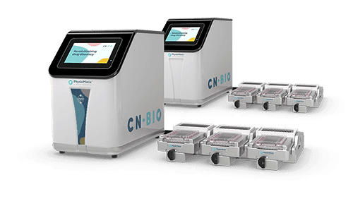
This webinar is based on our PhysioMimix Liver-on-a-chip system.
View our Q&A document from the live event.
Speaker Information:
 Dr Gareth Guenigault
 Dr Gareth Guenigault
Lead Scientist | Contract Research Services
CN Bio Innovations