
Resource > Webinars >
Addressing Unmet DILI Challenges: Novel insights from adaptable Liver microphysiological systems
Webinar Series 9 Episode 1
Filed under: ADME, DILI, and Safety toxicology

Video content if present
Drug-induced liver injury (DILI) remains a major challenge in drug development, especially when traditional models fail to capture complex or species-specific risks. Addressing these unmet workflow limitations is essential for improving confidence in drug safety assessment, particularly for multifaceted liabilities such as DILI.
In this webinar, we will demonstrates how Liver microphysiological systems (MPS) can be adapted to investigate different areas of toxicology – from lead optimization screening to identifying and understanding species-specific risks, informing study design and supporting mechanistic investigations of adverse DILI events in humans.
Drawing on recent peer‑reviewed studies, we will present a case example showing how Liver MPS studies compare across human and preclinical animal species. We will also explore challenges in evaluating cholestatic drug effects that cause DILI. Using a side-by-side comparison of multiple Liver MPS configurations, we will highlight how biological fidelity varies across systems and what is required for robust and reliable mechanistic cholestasis research.
Attendees will gain insight into where MPS adds the most value for reducing translational risk and increasing confidence in human safety outcomes.
Key Learning on how Liver MPS are used to address unmet DILI challenges:
See how to adopt and adapt MPS for different areas of toxicology and safety testing
Understand the importance of using MPS for cross-species DILI comparisons
Learn how MPS enables more complex and latent DILI effects to be unlocked
Explore what’s required of an MPS to evaluate cholestatic drug effects
View our Q&A document from the live event.
Webinar Transcript
00:00:21: Hello and welcome to the latest Technology Networks webinar, addressing drug-induced liver injury (DILI) using adaptable liver model systems.
00:00:28: I’m your moderator, Katie Brighton, Science and Newsletter Writer for Technology Networks, and I’m excited to be here to host today’s session.
00:00:35: We have a fantastic presenter, Dr. Emily Richardson, who will be sharing valuable insights with us. Emily is a Biology Group Leader at CN Bio, where she oversees the development and validation of microphysiological systems (MPS) for toxicology and safety assessment. She played a central role in creating the company’s [PhysioMimix] Lung and Lung/Liver MPS models, advancing their use in infectious disease research and the evaluation of inhaled therapeutics. Her expertise spans complex cell biology and real-world drug discovery.
00:01:07: She received her degree in Biochemistry and Molecular Medicine from the University of Nottingham and a PhD from the University of Leicester, where her work in 3D cell culture focused on mechanisms driving highly metastatic lung cancers and continues to shape her approach to developing more predictive and robust human-relevant models.
00:01:26: [Q&A submissions now closed] After the presentation, we’ll have a Q&A session, and we encourage you to submit your questions at any time during the presentation. To do so, simply type your question into the box on the right-hand side of your screen and click send. We’ll do our best to address as many questions as possible within the time available. If you encounter any technical difficulties during the webinar, please click the chat box on the right-hand side of your screen to request assistance from our support team.
00:01:49: Without any further ado, I’ll now hand over to our speaker, Dr. Emily Richardson. Over to you, Emily.
Drug-induced liver injury (DILI): High impact on success
00:01:58: Hey, thank you very much for that introduction. I’m looking forward to talking to you today about Liver microphysiological systems and how they can be used to address unmet DILI challenges.
00:02:11: First of all, what is DILI? We’re talking about drug-induced liver injury here.
00:02:17: This historically has a really high impact on the success of drug candidates moving forward in clinical trials. About a third of drugs fail in clinical trials due to toxicity, and around 18% of that is due to hepatotoxicity.
00:02:36: You can see some examples here of some very recent fails in clinical trials due to DILI events. Obviously, these fails are hugely impactful on both finance and reputation, but more importantly, on the future access to treatments by patients. So, it’s really important that we tackle this.
00:02:59: But why is DILI so complex to predict? Well, it comes down to the liver being a very important and also a complex organ. Drugs, when they’re ingested or applied in the blood, tend to always go through the liver. They may accumulate in the liver. They are then often metabolized or altered in some way before being excreted.
DILI: A complex beast
00:03:31: Now, DILI can come about in various different ways. It can be directly from the drug, where the drug impacts the liver directly through alteration of processes such as bile acid dysregulation, which is called cholestasis, perhaps on different metabolism effects on enzymes or transporters; and also effects on different organelles such as mitochondria or ER [endoplasmic reticulum], which can result in oxidative stress and death of cells.
00:04:04: The metabolized drug can also cause toxicity, so formation of toxic metabolites from otherwise a safe drug is quite a common mechanism of DILI. Also, one that is increasingly a challenge in drug discovery is toxicity via immune interaction with the liver – so, immune mediated DILI.
00:04:27: This is particularly becoming a challenge with new modality drugs such as antibodies or cell therapies, where these activate the immune system, which can then damage the liver. So, DILI, because it is so complex, has been classed into namely three different classes.
00:04:49: Firstly, intrinsic being the most predictable form of DILI. This is often dose dependent, often has early onset. So, you see it very early after dosing. And this sort of DILI is often captured by standard assays, such as classic in vitro 2D hepatocyte cultures or in animals as well.
00:05:13: However, it becomes more complex when we start to consider indirect or idiosyncratic DILI, where these are somewhat unpredictable and they are often latent in onset. So, you need a fair amount of time before seeing any toxic events.
00:05:30: Indirect DILI tends to be related to the pharmacodynamics of the drug, whereas idiosyncratic DILI is much more phenotype driven through genetics, disease profiles, and individuals on the whole.
00:05:44: So, when we think about these two more complex DILI events, we need model that maintains all the key cell types in the liver that may be affected by adverse events. We need a model that has functional longevity to capture these latent onsets, onsets that we see in more complex DILI events. We need a model that has human metabolic profiles to understand this pharmacodynamics. And, we also need a liver model that can deliver mechanistic insights and also clinically relevant insights to allow us to better translate to the clinic.
Why is DILI so challenging to predict?
00:06:26: When we look at our current models, starting with animal models, these can be very useful. It’s a whole system. It really allows us to understand various different types of toxicity and interactions across the body.
00:06:43: Animal models obviously can maintain key cell types. They have all the different cell types there and also has functional longevity. We can do long-term studies on these animals, but ultimately they’re not human. They don’t have the same metabolic profiles, the same biology, the same immune systems. This makes it really challenging when we want to then translate to the clinic.
00:07:06: When we then consider in vitro models, such as a spheroid model, these can be very useful. They have very good throughput. We can do very high throughput assays with them. They can maintain key cell types. You can include co-cultures.
00:07:23: Some in vitro models have functional longevity, and some have human metabolic profiles. What I mean by that is that there is a huge range of different in vitro liver models currently available.
00:07:38: You can see an example of this from a study that was done by this group looking at various different liver cell models and their metabolic profiles. And what you can see here is that they use primary human hepatocytes as the standard. These were freshly isolated and they looked at the metabolic profile formation rate. You can see that they also compared this to various different types of liver cultures from cell lines, such as Hep G2 and HepaRG to also iPSC derived hepatocytes. You can see there’s a huge range in terms of the metabolic profiles of these, this is in a log scale. So, there really is a huge amount of variation across there.
00:08:35: The other challenge with some in vitro models is that often these have fairly limited longevity and also fairly limited cell numbers, which means deriving mechanistic detail from them can be very challenging without pooling various different models together.
Why are in vitro liver models failing?
00:08:55: You can see this here in this graph that was taken from this paper from 2020 by Rubiano et al., where they looked at various different liver models.
00:09:06: And here you can see spheroids, although they have some functional longevity compared to other models, they have fairly low (this is) metabolism here. Whereas, models such as sandwich cultures start high and then tend to decrease very quickly over time.
00:09:26: Why is this the case with the liver? The main thing it comes down to is that the liver has and hepatocytes have high metabolic and respiratory function. This means they need high nutrient and oxygen requirements. When they don’t have those requirements, often they de-differentiate and so can no longer be used as a good cell model.
How to stop in vitro liver models failing?
00:09:52: So, how can this be stopped? How can we stop these liver models from failing? We looked at this and our approach is to use primary human hepatocytes that are freshly taken from patients. We can look at patient to patient variation in terms of metabolism, ensuring we have good access to nutrients and oxygen. So, by doing, by circulating the media around cultures, we can induce mixing of these different variables and avoid gradients.
00:10:33: The final thing is to have a very precise engineered microenvironment for the liver cells to sit in that makes them think they are back in the liver microenvironment. This essentially can halt de-differentiation and maintain the cells in a more human physiological state.
PhysioMimix Liver microphysiological systems: In vitro models with unrivalled in vivo relevance
PhysioMimix Liver microphysiological systems (MPS)
00:10:57:Here I’m going to introduce you to the PhysioMimix® Liver microphysiological system. I’m going to refer to this as MPS throughout this presentation. These systems can also be known as Liver-on-a-chip or OOC [Organ-on-a-chip] is another term that is often used.
00:11:16: This is a schematic of our Liver MPS. We have an engineered liver scaffold that sits within the well. And the primary human cells are inserted onto the scaffold. This can be primary human hepatocytes (PHH) on their own if looking at things like metabolism. Other non-parenchymal cells can be added in, such as Kupfer cells or stellate cells, if you want to look at things like disease profiles and also drug-induced liver injury (DILI).
00:11:52: Within our Liver microphysiological system, we have an individual micro pump per well, which pumps and moves the media up and through the liver microtissues, circulating the media around the well and also inducing shear stress on the cells.
00:12:12: What you can see is the way that we’ve engineered this platform is to have a large surface area on the top to allow optimal oxygenation of the media, which again really benefits the cells.
00:12:28: You can see here in another schematic that the cells essentially sit within the micropores of the liver scaffold and they essentially form structures very similar to the liver sinusoid. The fluidic flow moves up and through these microtissues, and this allows optimal oxygen gradients to be maintained. This is what our liver plates look like.
PhysioMimix Liver MPS: Liver-12 plate & Liver-48 plate
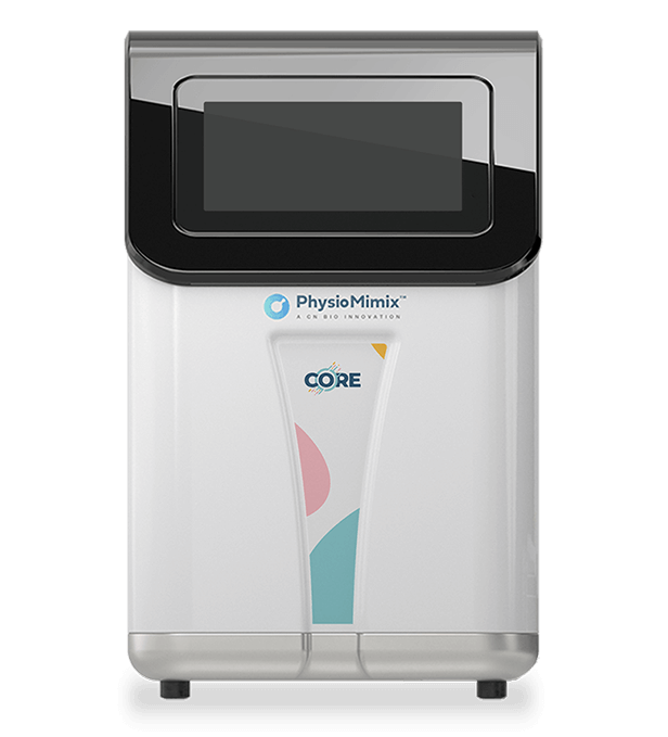
00:12:56: We have two forms of the plates. These plates can both be powered by the PhysioMimix Core Controller. This Controller sits outside of an incubator and feeds in through an Umbilical Cord, which attaches to the Docking Stations, which sit on the shelves of a standard incubator.
00:13:17: You can run up to six plates per [PhysioMimix] Core Controller, which allows a good amount of throughput, which I will go into a little bit more now.
PhysioMimix Human DILI: Scalable to requirements
00:13:28: The Liver-12 plate is our original Liver MPS plate. It has 12 individual chips, which you can see here. Within each of these wells at the end of the plate, you have your scaffolds where there are 301 individual micropores where the cells sit and the schematic that I showed you in the previous slide. This has been a really successful plate. It’s been very well qualified and used within various different contexts of use.
00:14:06: The feedback that we got from the field was, this is great, but we want more and we want more throughput. So, our engineers worked very, very hard to essentially make this plate a quarter of the size so that we can have 48 individual Liver-on-a-chip wells; and each of these run individually.
00:14:30: You can see that the Liver [Liver-48]micro scaffold is a quarter of the size of the Liver-12 scaffold. What this means is that we can run even more chips per PhysioMimix Core, so 288 chips compared to already quite a good throughput of 72 chips in with the Liver-12.
00:14:51: What this means is that you have a reduced chip size, so less recoverable media. This is, again, is essentially a quarter of the Liver-12 plate. What we’ve managed to maintain is the ratio between the media and the cells, which means that you’re still able to access the complexity of the cell culture, maintain that complexity, and also be able to still measure important clinical markers such as ALT and albumin.
00:15:20: In the Liver-12, because it has a much higher recoverable media volume and larger amounts of cells, you can do even more analysis to get the deepest insights into various different mechanisms. So, this is what biologically the liver MPS looks like.
PhysioMimix Liver MPS
00:15:41: When we start the cultures, we add our primary cells in a single cell suspension at the beginning of the experiment.
00:15:50: Over several days, they mature into these micro tissues, which look a little bit like an American style doughnut shape. You can see here when we characterise these using various different methods, such as IF staining, that. They produce albumin, they form bile canaliculi, which we’ll go into a bit further in this presentation. They’re polarised and form tight junctions, which you can also see in this beautiful IM image here, and also the formation of bile canaliculi, as you can see in this image. Importantly, when we look at these models, we’re able to capture metabolism over time. So, they have this very long functional longevity.
00:16:38: Here, this is taken from the same study I showed earlier, where it’s a collaboration with the FDA, where they’re analysing the Liver MPS for use in drug safety assessment. You can see here that the Liver MPS is able to maintain metabolic activity over time. We’re also able to look at the metabolic profiles of different hepatocyte donors. Each of these are different donors available commercially. 00:17:11: We’re able to look at the various different metabolic activities of different enzymes. And importantly, we validate ourselves internally from commercial sources, which allow good availability for use by other customers.
PhysioMimix enhances safety studies
00:17:31: When we’re thinking about safety, there is obviously a long list of safety concerns and tests that are done over the drug development and discovery cycle.
00:17:44: At the beginning, when you have a huge amount of different candidates, you want to do some very high throughput screening, you want to be using high throughput methods. Using your in silico methods, first of all, going into your 2D, perhaps spheroid screening cultures, looking at overall toxicity, looking at things like ATP and LDH to get an idea of the sort of overall DILI risk; and potentially narrowing down clinical concentrations to be tested.
00:18:15: At that point, this is a great time to use a human DILI assay because what you’re able to look at is much more detail in terms of the risk and also looking at detailed analysis of concentrations. That really allows you to gain even more confidence before proceeding with lead candidates before going into non-clinical tests. It also allows you to better inform the design of those tests as well and potentially avoid delays.
00:18:50: The other assay which we have recently developed is a cross-species DILI assay. The idea of this is to essentially look at the differences in species-specific toxicities. Because we know that in non-clinical trials we have to use a two animal species, but often it’s very challenging to know which of these species are going to be most predictive. So, by running animal MPS prior to this non-clinical, very expensive, IND-enabling study, you can look at what those risks could be in an MPS and compare it to human. Importantly, what that allows you to do is gain more confidence again before proceeding.
00:19:39: We also have customers coming to us after non-clinical studies, where they have potentially seen two different responses, maybe toxicity in a dog, but not in the rat. Which one is correct? By using both of those models plus a human model in the MPS, it gives you more confidence as to what the human output is going to be when you get to those clinical trials.
00:20:06: And then finally, the other way in which MPS can be really useful is looking very deeply into mechanisms of toxicity. So, using it for investigative toxicology studies. What this does is to really allow you to map any toxic events that may have happened potentially in the clinic or before that; and also, allows you to avoid these adverse events in the future.
PhysioMimix DILI assays & case studies: Predict complex & cross-species DILI with sensitive in vitro hepatotoxicity assays
PhysioMimix Human DILI assays
00:20:38: I’m going to start just by showing you our standard human DILI assay. The overall timeline of what this looks like is very similar between the Liver-12 and the Liver-48. The first step is to prime these plates to make sure that all of the components are wetted within the plates.
00:21:01: On day zero, we seed hepatocytes and Kupffer cells together. We include Kupffer cells as the immune component of the liver, the tissue resident macrophages in the liver as this is a really important part of any potential DILI response.
00:21:20: After four days of culture, we then do a liver quality control test where we look at important health and functionality markers to ensure that all chips are functioning, robust, and are within the same remit; and then we can start dosing and sampling.
00:21:40: Because our plates are open well formats, we can dose and sample as much as is required and as much as is applicable to the drug candidate that is being tested.
00:21:53: We take those samples. The shortest amount of time we will dose for is four days, but we can go up to 28 days as an example. I know examples of where people have gone beyond that testing but at that end point, we can then take out the tissues and the rest of the media and we can further analyze the tissues. We can look at them under the microscope to look at structural changes. We can also do high content analysis, such as omics analysis, flow cytometry, or microscopy as examples.
00:22:33: We’ve qualified this model very thoroughly in the past. First of all, we used tool compounds set out by the IQMPS consortium, which is taken from the Baudi et al. paper from 2019. We applied these drugs in various concentrations to the liver model and measured various outputs such as albumin, urea, ALT and AST, and ATP. And we graphed these to the margin of safety of each of these outputs. What you can see here in this graph really nicely is that we’re able to characterise these drugs from low DILI concern to high DILI concern. And this really nicely matches what is known of these tool compounds from previous clinical outputs.
PhysioMimix DILI assay: qualified using IQ MPS Consortium tool compounds
PhysioMimix predicts DILI risk of tool compounds
00:23:31: To show you an example of what this sort of data can look like, I use the example of troglitazone and pioglitazone. It’s very well known, troglitazone being a very highly toxic drug, a DILI rank eight. It was withdrawn from the market following release after various liver failure related deaths. And pioglitazone is the safe equivalent that is still used in the clinic. What you can see here in yellow is that troglitazone reduced the functionality of the liver microtissues with increasing concentration and increased liver stress markers like ALT and AST, and reduced the overall ATP content. This shows really nicely that we’re able to match the hepatotoxic risk to what is known for the clinic. That’s kind of the first stage when we’re looking at safety screening.
PhysioMimix enhances safety studies – The translation of MPS
00:24:38: I’m now going to talk about species translation studies. The ideal when we are making MPS models is that we make human MPS models to predict the human outcome. That is what we want to know. We want to make sure that these drugs are safe in a human. However, comparing human in vitro data to clinical data can be very challenging. There’s often limited available clinical data and there is a lot of convincing that people need from this data. So, something that has been discussed widely across the field has been animal MPS. And the reason behind this is that we have a lot of animal data which can be directly compared.
00:25:33: So, if we develop an animal MPS, and we test drugs that have been tested in vivo and we get the same outcome, it gives us more confidence that our human MPS is able to predict our human in vivo.
00:25:51: This has been discussed, you can see here from these two opinion articles by the FDA and also the IQMPS consortium. The animal MPS has also been suggested as a very useful tool in other areas as well, such as veterinary medicine.
Adapting PhysioMimix to enhance translatability: Cross-species DILI assay
00:26:10:We set out recently to develop these models. We developed a dog and a rat Liver MPS. They’re used in almost exact same way as a human MPS. So, we see the hepatocytes, we QC, and then we dose and then we take down the tissues.
Tolcapone hepatotoxicity identifies in rat in vivo studies
00:26:34: To show you an example of what this sort of dataset looks like, I’ll use the two tool compounds, tolcapone and entacapone. Both of these were used as therapies for Parkinson’s disease. However, tolcapone is a DILI rank 8, as it was associated with several instances of DILI. Entacapone is broadly thought of as safe. However, there has been association to low increase of ALT in the clinic. Importantly, the risk of tolcapone wasn’t detected in the non-clinical studies, but there have been various in vivo studies since this that have been able to identify it in both the dog and the rat.
Tolcapone hepatotoxicity detected across species in Liver MPS
00:27:19: When we apply these drugs to our cross-species Liver MPS, what we can see with tolcapone is that each of the animal species were able to detect the toxicity of tolcapone in slightly different ways. But we saw increases in ALT and decreases in albumin as an example of the entire data set. We’re able to capture EC50 values from the human and the rat, but not the dog. And you can see that the rat was somewhat more sensitive in the albumin. Interestingly, what we see with Entacapone is that there are no changes in albumin across each of the three species. However, we are able to capture an EC50 from the human of the ALT increase with entacapone, so at a higher concentration, which nicely matches what was seen in clinical trials.
TEX-VAL case studies
00:28:24: I also want to bring in a case study of a recent publication from the Tex-Val Consortium. The Tex-Val Consortium is a consortium made-up by the Texas A&M University group that have various other stakeholders included from various drug developers to the NTP. The aim of this consortium is to test the feasibility of technology transfer from MPS developers, like ourselves at CN Bio to potential end users. They do some really fantastic work looking in depth at different MPS platforms and how they can be used, adapted, scaled to requirements from drug developers.
Cross-species MPS: TEX-VAL case study 1
00:29:21: The first case study I’m going to show you is a publication from last year, where the group looked at adapting the system to four different species – human, monkey, dog, and rat.
This paper was actually picked up by Science, where it’s quoted as being “one of the best papers they’ve seen in this area.” So, I would highly recommend reading this paper if you haven’t already.
What was seen when the group adapted the MPS to these different species, so human, monkey, rat, and dog, they tested the hepatocytes in a 96 well plate and in the PhysioMimix Liver-12.
00:30:14: What they saw was that for human and monkey, so these top two graphs,(here, I’m just showing the albumin results, but in the paper, they also look at other functionality markers as well)there was an enhanced longevity of albumin secretion, also increased albumin secretion over time compared to 2D.
In rat and dog, it was somewhat comparable to 2D, with a bit more enhanced variability in the rat and also lower amounts of albumin produced in the dog. which is also something that we have seen in our internal assays and something we’re continuing to work on with cell providers.
This is an important part of consideration of MPS development, really considering the species cell numbers, how they are in vivo, and what to expect in terms of outputs.
00:31:13: What the group then did was to really go into depth into looking at the interspecies difference in DILI outcomes. They looked at three specific drugs – chlorpromazine, bosentan, and fialuridine. This graph here shows a nice overall summary of the outputs from standard DILI markers, ALT, AST, LDH, albumin, and urea.
00:31:44: This was looking over time. They also had other graphs looking at concentration dependent changes. What you can see is the fold differences from the vehicle controls compared across the different animal species. What you can see is that there are differences and also some similarities depending on the different drugs.
00:32:06: The group went even further and upped the takedown at day 10 of dosing. They took the liver microtissues and used them for transcriptomics. So, what you can see here in A is that all of the drugs cause significant gene alterations in the human in blue. Very small differences in the monkey. Some changes in the rat for chlorpromazine and for fialuridine, and also in the dog for fialuridine. What was really interesting in the study is that they also looked very closely into the overlap of different drug effects.
00:32:52: Here I’ve used the example of chlorpromazine from the paper, where they were looking at how gene changes overlap between the different species. What you can see here is that in the case of chlorpromazine, the human and the rat were the most similar in terms of the gene changes. This was found mainly in protein and RNA metabolism pathway changes.
Despite the monkey having a low amount of genes dysregulated, what they found that was around half of the genes that were altered were common between the human and the rat and the monkey.
PhysioMimix enhances safety studies
00:33:32: This study was particularly interesting because not only did it show the adaptability of the system to culture and test different species, but also the ability to scale the analysis to really get a deep mechanistic insight into the toxicity. That’s what we’re going to go into a bit further now, looking into how the system can be used in investigative toxicology studies.
Limitless end-point analysis from each well of PhysioMimix Liver Plates
00:34:11: As mentioned previously, in the Liver-12, there is a large amount of media volume and microtissue to analyze. Because of this, we can look at various different outputs from sampling. So, from the media, you can look at various different mechanisms of DILI – from things like cholestasis, mitochondrial dysfunction, to things like steatosis and fibrosis.
From the liver microtissues, as mentioned previously, you can use these to do things like various different omics analyzes, and also high content microscopy and things like flow cytometry as well.
Common mechanisms of DILI
00:34:59: The common mechanisms of DILI are shown here. This is not necessarily all of them, but certainly some of them. As mentioned previously, reactive metabolites are one of the key mechanisms of DILI. These can cause protein abducts, causing increased inflammation, immune DILI interactions as well, and damage to the cell directly. This can cause things like oxidative stress by dysregulation of mitochondria. This increases reactive oxygen species, which can directly damage the cell, but again, also cause inflammation. Damage of the mitochondria can also cause an increase in free fatty acids. The drug can also dysregulate the transport of fat, which can cause steatosis or lipid accumulation in the cells. Stellate cells can also be activated through damage, which can cause increased fibrosis. So altogether, this kind of goes towards a MASH phenotype of which we also have a very robust model in the PhysioMimix system.
Cholestasis: TEX-VAL case study 2
00:36:23: Cholestasis, as I’ve mentioned before, is a fairly common DILI mechanism, whereby there is accumulation or dysregulation of bile acids that can be through inhibition of transporters and also metabolism effects as well.
00:36:43: I’m going to focus on how we’re able to look at cholestasis, first of all. And again, I’m going to use a case study from the Tex-Val Consortium.
00:36:55: A paper published last year looking at various different Liver MPS systems and configurations, particularly in how they can capture cholestatic events. This was a really interesting paper where the authors used various different cell types – from cell line HepaRG, to primary human hepatocytes, and also iPSC-derived hepatocytes in three different formats – in a 96 well plate, in PhysioMimix Liver-12, and in the two or three lane Organoplate from Mimetas.
00:37:40: What we’ve shown initially in the study looking at functionality in a seven day study on primary human hepatocytes and the iHeps over seven days and over 28 days in Hepa-RGs. You can see that there is maintained functionality across the Liver chip in albumin, urea, and CYP over each of the time points; fairly similar to 2D.
The authors then went further to look into metabolic activity. Obviously, this is an important part of hepatocyte function and also the mechanisms of DILI. They looked at a seven and a 30-day treatment of a compound called bosentan, which is a known BCEP inhibitor causing cholestasis. It’s also a CYP3A4 inducer as well. Here, you can see the CYP activity was monitored across the 96-well plate, Liver-12, Organoplates in HepaRG and PHH cultures.
00:38:54: What you can see here highlighted in the Liver-12 is that the metabolic baseline function in the control in CON remained high.
00:39:08: When bosentan was applied, you could see enhancement or induction of that activity, both in HepaRG and also in PHH at both seven days and at 30 days in the HepaRG. The team then went on to look at bile acid production, obviously key when thinking about cholestasis.
00:39:35: What you can see here again in seven and 30 days treatment, this time using three different drugs, was that both HepaRG and PHH produced bile acids in the Liver-12. The PHH overall produced more bile acids than HepaRG and also produced more glycine-conjugated bile acids, which are much more human liver like.
00:40:05: What you can see in this figure is that the Liver-12 was the only model able to capture the decrease in bile acid upon treatment with drugs.
00:40:16: Both in HepaRG, but also more significantly in the PHH cultures, you see this reduction in bile acid production in the media. This is really important in terms of being able to capture cholestasis, but also considering how you model these different events in line with your context of use. This is a really important factor that is outlined in this publication. I would highly recommend looking further into this one.
Detecting cholestasis with transcriptomics
00:40:59: Continuing with the theme of cholestasis, we have been doing some further work at CN Bio to understand how we can continue to push the understanding of this using our system. For this, we used three tool compounds – diclofenac, which is a DILI rank 8, but still used in the clinic, but has a black box warning, and then troglitazone and pioglitazone, which I introduced earlier.
00:41:15: Diclofenac and troglitazone have both previously been shown to have cholestatic events. So, we wanted to look further into this as well as other mechanisms. We tested these drugs in the system over 10 days of dosing at a clinical and 1.5 times clinical dose. Samples were taken every two days. At the end of the study at day 10, we took the cell lysates for transcriptomic analysis. This work was done by Dr. Ovidiu Novak in my team.
00:42:02:What you can see here, when we look at the bile acid biosynthesis process, so metabolic enzymes and also transporters involved in bile acid transport in and out of the cell, we can see that troglitazone and diclofenac both negatively impacted the expression of these key processes, which is in line with their cholestatic nature.
Detecting oxidative stress with advanced clinical biomarkers: Diclofenac
00:42:34: We then looked further into other mechanisms. This isn’t necessarily showing everything from this publication that is currently being prepared; a snapshot of some other markers we looked at.
00:42:47: We were keen to understand more about oxidative stress and resulting cell death. So, we tested several markers identified from the Predictive Safety Testing Consortium that were identified as potential beneficial daily biomarker candidates – we used ARG1, MDH1, and SDH1. What you can see here that is really nice in diclofenac is that at the clinical concentration using this donor, we didn’t see much effect of the drug to any sort of oxidative stress outcomes, apart from an increase in MDH1.
00:43:33: When we then increase that concentration to 1.5x the clinical concentration, we start to see these sensitive biomarkers increase around four days of dosing. Some of them being a little bit more early, a bit earlier and more sensitive than that.
00:43:53: Corresponding with that, what we can then look at are cell death markers. Here we use cytokeratin-18 for the necrosis and cleaved cytokeratin-18 for apoptosis, and you can see that we’re able to capture the apoptosis that then occurs at the 1.5x clinical concentration, as well as some necrosis that also follows.
00:44:16: What this really demonstrates is that this assay is sensitive to concentration dependent toxicity and can capture the mechanisms and timing of the adverse events. So, look out for this publication coming soon because we’ve got even more data to come on that.
PhysioMimix enhances safety studies
00:44:38: To summarize, this is how we use our PhysioMimix DILI assay within safety studies. You can use it to safety screen using the human DILI. You can look at species translation studies to understand species-specific toxicity, and also use it for investigative toxicology studies as I’ve just shown.
Reasons to choose the PhysioMimix Core DILI assay
00:45:02: And together, why it’s useful to use these MPS assays and specifically the PhysioMimix Core DILI assay is that these assays have been shown again and again to be highly reliable and robust, with low variance between wells. They’re adaptable and scalable to your context of use.
00:45:24: They are used to test new drug modalities. I haven’t shown this data today due to time, but it’s a really good model for looking at those human relevant paths where there are no available alternatives, particularly with animal models.
00:45:40: And finally, as I hope I’ve shown today, you’re also able to capture mechanistic and clinical translatable insights.
3RsC-FDA DILI project
00:45:53: The final thing I want to highlight is something we’re very excited about. We are a part of the 3RsC-FDA cross-platform DILI project, which has recently been accepted into the FDA ISTAND program.
00:45:58: We’re one of the eight commercial MPS providers. We have already tested eight blinded DILI tool compounds. The unblinding has just happened. We’re very excited about the results. I’m not going to say anything yet but continue to watch this space.
Let’s Change the game, together
00:46:44: This is a really good example of how there is this huge regulatory backing for MPS now, both with new regulations aiming to phase out and reduce animal testing, and also this enhanced awareness that MPS data is really translatable. And so, MPS technology is really becoming an integral part of drug discovery and development.
00:46:52: And if you aren’t already using these models, now is the perfect time to start understanding and using them within your drug development and discovery.
PhysioMimix Core: The only MPS that’s easy to adopt, adapt, and scale right away!
00:47:05: To finish off the PhysioMimix Core, hopefully today I’ve been able to show you how easy it is to adopt, adapt, and scale to your context of use. The system is open well [format] and familiar to any biologist.
00:47:21: With the complex engineering that people are often worried about with these complex systems, they are integrated within the system. So, all you have to do is think about the biology in front of you.
00:47:32: You can adapt the system easily for different organs, cell types, timelines, different drug modalities, and also scale it to your throughput requirements.
How to access our MPS technology
00:47:44: If you want to access the MPS technology, you can either purchase the system to use in your own lab, or you can access the technology through our Contract research services (CRS) using our Organ-on-a-chip experts at our HQ in Cambridge, UK.
00:48:38: Please feel free to get in touch if you’d like to know any further information. I’ll finish there. Thank you ever so much for your attention today. If you want any further information, please contact me. My e-mail address is there. I will welcome any questions now. Thank you.
Speaker

Dr Emily Richardson
Biology Group Leader
Throughout her time at CN Bio, Dr Richardson has led multiple collaborative and grant funded programmes and acts as a key liaison across academic partners, pharmaceutical organisations, contract research organisations, regulatory bodies, and standardisation groups.
Her expertise sits at the intersection of complex cell biology and real world drug discovery, informed by industry experience in cellular therapeutics and specialism in complex in vitro modelling. She received her degree in Biochemistry and Molecular Medicine from the University of Nottingham and PhD from the University of Leicester, where she used 3D cell culture to uncover molecular mechanisms driving highly metastatic lung cancers – expertise that continues to shape her approach to developing more predictive and robust human relevant models today.