How a partnership between the FDA and Organ-on-a-chip developers has the potential to fast-track drug development
From a small discovery in a research lab to a capsule on a pharmacist’s shelf – the path of drug development is a long one. Whilst scientists discover or engineer many new compounds and molecules every year, very few of them go on to become successful medicines.
Why does drug development take so much time?
When scientists and clinicians consider new drug treatments, they need to take into account two very important factors: safety and effectiveness. If a drug kills cancer cells in a dish, but it also kills healthy human cells, it would quickly fail drug safety tests. On the other hand, if another cancer drug left healthy human cells alone but also ignored cancer cells, it wouldn’t be a good cancer drug either. Of the tens of thousands of potential drugs that go through pre-clinical tests and clinical trials, unfortunately the majority fail due to concerns with safety, effectiveness, or some combination of the two.
Translatability, or how well a certain drug works in laboratory-based in vitro cell culture, in an animal model, and finally in a human, is certainly a contributing factor here. Sometimes, a drug may work fantastically well in human cells in a dish, but when it is then tested in humans in a clinical trial, it may have no effect. The inability to translate results from in vitro cell culture or animal models to actual human patients is a major roadblock in drug development.
To better mimic the physiological processes that occur organs in the human body, scientists and bioengineers have developed microphysiological systems (MPS), also known as Organ-on-a-Chip (OOC) systems. By incorporating the 3D architecture of a human organ together with all of its different cell types, plus intricate microfluidics that circulate medium through the tissues (similar to human blood flow), Organ-on-a-Chip systems offer enhanced human relevance over standard 2D in vitro systems and 3D spheroids.
If they are more physiologically relevant, why not use Organ-on-a-chip systems for all drug development studies?
The simple answer: this technology is still relatively new. Because these systems are on the cutting-edge, only a small but growing number of drug developers are using an organ-on-a-chip system and have tested how it works in their own hands. For a team to invest, they would want to ensure that any given solution generates data that does indeed translate into human outcomes and offers clear benefits over more established systems like 2D in vitro cell culture or animal models before making a commitment.
For this reason, the Center for Drug Evaluation and Research (CDER) at the Federal Drug Administration (FDA) partnered with a number of companies/institutes who develop OOC systems in order to test the systems and evaluate them directly in the context of drug development. If successful, this most interesting of collaborations has the potential to reduce the time it takes for a drug to be developed and approved for patient use, and in doing so, it will greatly reduce the costs of bringing these new drugs to patients in need.
How does this collaboration between the FDA and Organ-on-a-chip developers work?
The traditional path of drug development generally involves testing a potential drug in 2D in vitro cell culture models, animals, and then in humans. However, to see how good a particular OOC system is at predicting actual clinical outcomes, the FDA scientists start at the end of this process with published clinical data about a drug. In this way, the FDA formulates a hypothesis about how a particular drug works in a specific biological context, from which they can test their hypothesis using OOC system.
The FDA is particularly focused on liver and cardiac OOC systems because over 75% of drugs for liver and cardiac applications are withdrawn due to safety-related concerns. Additionally, problems with these drugs are often caught late in the drug development process. If OOC can better predict how well a drug will work in clinical trials, studies have estimated that they could save more than $100 million in drug development costs.
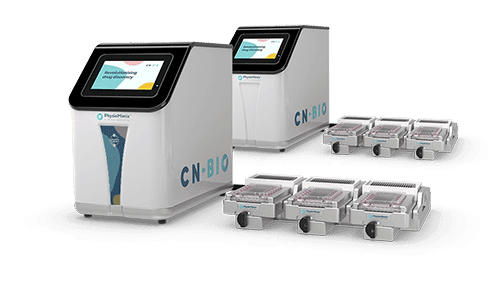
In particular, the development of good in vitro liver models is key for more efficient drug development. One major role of the liver is to metabolize or transform drugs facilitating their removal from the body.. Because of these functions, in vitro liver models are incredibly valuable for studying drug toxicity, metabolism, clearance, and interactions with other drugs and the body in general. Currently, the FDA is working with CN Bio and using our PhysioMimix® OOC Microphysiological System to evaluate an in vitro liver-on-a-chip model, sometimes referred to as a Liver MPS.
What are the results of the collaboration between the FDA and CN Bio with the PhysioMimix® Liver-on-a-chip model?
The PhysioMimix® liver-on-a-chip model mimics the physiological environment of the liver by incorporating the different cell types found in the liver – primary hepatocytes, hepatic stellate cells, and Kupffer cells – in a 3D environment under fluid flow. The presence of these different cell types allows scientists to investigate and profile human liver responses to different drugs. For example, if a drug caused an inflammatory response, hepatocytes and Kupffer cells would be affected, but, if the drug caused necrosis, then primary hepatocytes would die.
To understand how the Liver-on-a-chip model handles drug toxicity in the context of an inflammatory response, FDA scientists used it to test the effect of two drugs: FDA-approved levofloxacin and the liver-toxic drug, trovafloxacin. The FDA scientists found that the Liver-on-a-chip model was able to detect trovafloxacin-induced toxicity by reading out increased cell death. They also saw that their results were consistent when the test was conducted at different sites and with different batches of Kupffer cells – thus, lending them even greater confidence in the results. Additionally, by measuring albumin production from primary hepatocytes, the FDA scientists found that the Liver-on-a-chip model maintained its function longer and functioned better than the traditional 2D hepatocyte cultures and 3D spheroid culture methods. Therefore, by incorporating media flow through the 3D culture set up, this Liver-on-a-chip model offers clear performance advantages over other in vitro culture methods.
What Comes Next?
In the future, the Center for Drug Evaluation and Research at the FDA is planning to expand their OOC collaboration by evaluating other single-organ systems, such as a Kidney-on-a-Chip system and CN Bio’s Lung-on-a-Chip platform, and investigate the potential for multi-organ solutions to provide organ-organ crosstalk insights that currently cannot be revealed in vitro. Additionally, looking toward a future where medicine will be more personalized, the FDA hopes to take cells from patients, differentiate them into different cell types, and test how a specific patient’s cells react to a particular drug in OOC systems.
So, watch this space, with these almost life-like OOC platforms, scientists and regulators move another step closer to bringing life-saving drugs from the lab to your neighborhood pharmacy – in record time.
Author: Stephanie DeMarco
If you would like to hear this story direct from the regulators themselves, please tune into the demand webinar presented by Dr Alexandre J.S. Ribeiro (Staff Fellow (Biological Scientist)
FDA) entitled:
Alternatively, take a look out for the peer-reviewed publication in the Journal of Clinical and Translational Sciences


