

Resource > Blogs >
Cancer-on-a-chip melanoma model for CAR T cell therapy development in solid tumors
Filed under: Disease modeling, General OOC, and Oncology

Cancer-on-a-chip models are becoming a central tool for testing immunotherapies against solid tumors. By incorporating dynamic flow, chemokine signaling, and a human endothelial barrier, these models can recreate aspects of immune-cell trafficking and tumor-cell interactions that standard static cultures may miss. CAR T cell therapy reshaped the treatment of hematological malignancies but has stalled against solid tumors. This post looks at how the PhysioMimix® platform can be used to study melanoma, using a perfused melanoma-on-a-chip model built for CAR T cell testing as the worked example.
Why solid tumors resist CAR T cell therapy
CAR T cell therapy changed what is possible in hematological malignancies. Reaching solid tumors with the same approach has proven far harder. Solid tumors rarely present the clean, disease-specific surface antigens that make a leukemia cell an easy target, and they sit inside a microenvironment built to keep immune cells at bay. Hypoxia, dense extracellular matrix, disorganized and leaky vasculature, and a thicket of immunosuppressive signals all work against T cell infiltration. A CAR T cell that clears a patient’s leukemia may never reach, enter, or kill the cells inside a solid tumor, such as a melanoma nodule.
This is complicated by the challenges posed by melanoma specifically. It accounts for a small fraction of skin cancer cases but approximately 80% of skin cancer-associated deaths, and it carries one of the highest mutational burdens of any cancer. It is aggressive, often hard to catch early, and biologically unlike the tumors that most preclinical models were built around. That mutational burden is a large part of the reason, since it drives antigen variability from one patient’s tumor to the next, which is a hard problem for a therapy that depends on recognizing a single surface target. For therapy developers, meaningful evaluation must establish whether a CAR T cell can traffic to the tumor, infiltrate its microenvironment, persist, and retain its cytotoxic activity.
Recreating the vascular journey to the tumor
An infused CAR T cell circulates in the bloodstream, migrates across the vessel wall that has been activated by inflammatory signals, infiltrates the tumor, and finally kills cancer cells by cytolysis. Each step is a checkpoint where a drug can fail. A T cell can circulate perfectly well and still fail to cross an activated endothelium. It can cross and still lose potency by the time it reaches the tumor.
Most in vitro models capture one slice of this. Static co-culture, where T cells and tumor cells are mixed together in a well, measures killing but skips everything that comes before it. The T cells never circulate, never meet a vascular barrier, and never have to earn their way into the tumor. Animal models capture more of the whole, but mouse biology diverges from human biology in ways that matter for this disease. Mouse skin differs in architecture and in the ratio of melanoma to non-melanoma cells, so a mouse cannot be tuned to represent a human melanoma with much precision. The result is a translation gap. Findings that look convincing in a simplified system or a mismatched animal do not transfer cleanly into patients, and this can result in drug testing failures.
This is the space that MPS-based models, and specifically perfused tumor-on-a-chip models, are built to fill. A cancer model assessed on the PhysioMimix platform can recapitulate the steps a static assay loses, without adding complexity for its own sake. The field has no shortage of tumor chips, and many are narrow, built to answer one specific question and hard to reuse. The task is to include the steps that change the answer and leave out the ones that do not.
Building a melanoma-on-a-chip on the PhysioMimix platform
PhD student Daniel Rieger built a perfused melanoma-on-a-chip model on the PhysioMimix Core System. The platform circulates culture medium to model blood flow and shear stress, which is the feature that lets the whole cascade play out rather than just its final step. This work is part of Melomanes, an EU-wide consortium developing an immunotherapy that pairs anti-HLA-G CAR T cells with magnetic nanoparticles capable of inducing localized hyperthermia. The CAR T cells target HLA-G, an antigen expressed by melanoma. In healthy tissue, HLA-G is largely confined to the extravillous trophoblast at the maternal-fetal interface, where it suppresses NK and T cell cytotoxicity so the maternal immune system does not reject a semi-allogeneic fetus. Melanoma co-opts that tolerance signal to evade immune surveillance, and the molecule the tumor uses to hide becomes the handle the CAR T cell grabs. Its restricted distribution in healthy tissue is also what limits the risk of on-target off-tumor toxicity.
The model uses a Transwell-based, dual-compartment design. Human umbilical vein endothelial cells (HUVECs) are seeded on collagen type IV-coated membranes and cultured under perfusion, forming an endothelial barrier that stands in for the vessel wall. On the tumor side, SK-MEL-5 melanoma cells are engineered to express HLA-G stably, giving the CAR T cells a target. The model uses an immortalized cell line rather than patient-derived tumor material, which keeps antigen expression controlled. Anti-HLA-G CAR T cells are introduced into the system and, guided by a chemokine gradient set with CXCL10 (IP-10), migrate across the endothelial layer toward the tumor, where their killing can be measured.
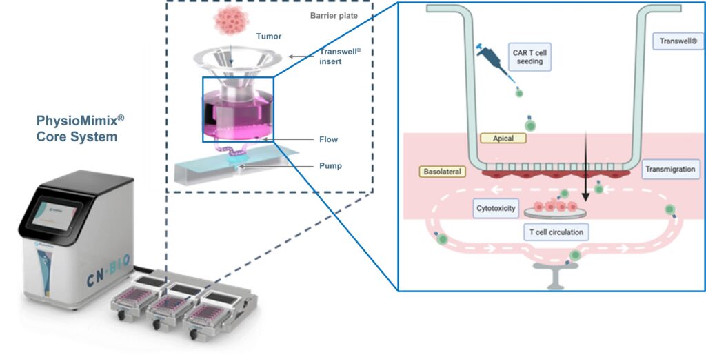
Each piece maps onto a step of the cascade. The perfused HUVEC barrier is the vessel wall the T cells must cross. The chemokine gradient is the recruitment signal that pulls them across. The HLA-G positive SK-MEL-5 cells, grown on collagen-coated discs, are the target they have to reach and kill. The readouts are chosen to match. Barrier formation is tracked by immunofluorescence staining for F-actin and the tight-junction protein ZO-1, by transendothelial electrical resistance (TEER), and by paracellular permeability using FITC-dextran. Migrating T cells are quantified with CellTiter-Glo and automated cell counting, and cancer cell death is read out by bioluminescence from luciferase-labeled tumor cells. Because the compartments are separate, each step can be measured on its own and tuned independently.
What the melanoma-on-a-chip model showed
Building the barrier turned out to depend on conditions a developer can control. Endothelial cells seeded on the collagen type IV-coated membranes formed tight barriers, and both perfusion and the collagen coating improved barrier formation compared with static conditions. Permeability and TEER assays showed that barrier integrity peaked after 48 h of perfusion.
The barrier also responds to inflammation the way a real vessel does. Treating the endothelium with TNF-alpha drove VCAM-1 upregulation, loss of cell-cell adhesion, changes in cell shape, and increased permeability. That matters because the activation state of the endothelium is not cosmetic. CAR T cell transmigration across the barrier depended on the endothelial activation state, on the chemokine concentration, and on perfusion. Change the vessel wall, and you change how many T cells get through.
Cancer cell death was governed by the effector-to-target ratio, by perfusion, and by whether the tumor actually expressed the target antigen. The finding that carries the most weight for therapy development is this: CAR T cells retained their cytotoxicity after crossing the barrier, but they killed less efficiently than T cells placed in direct contact with the tumor. The barrier itself blunts efficacy. A static assay, by design, can never show you that, because it removes the barrier before the experiment starts. For a developer, that reframes the migration step as something to engineer rather than assume, and it flags when a candidate that looks strong in a dish may lose ground on the way to the tumor.
Taken together, the model recapitulated key aspects of the CAR T immunological cascade in a single dynamic system, covering circulation, transendothelial migration, and tumor cell killing, using primary anti-HLA-G CAR T cells alongside human endothelial and cancer cells.
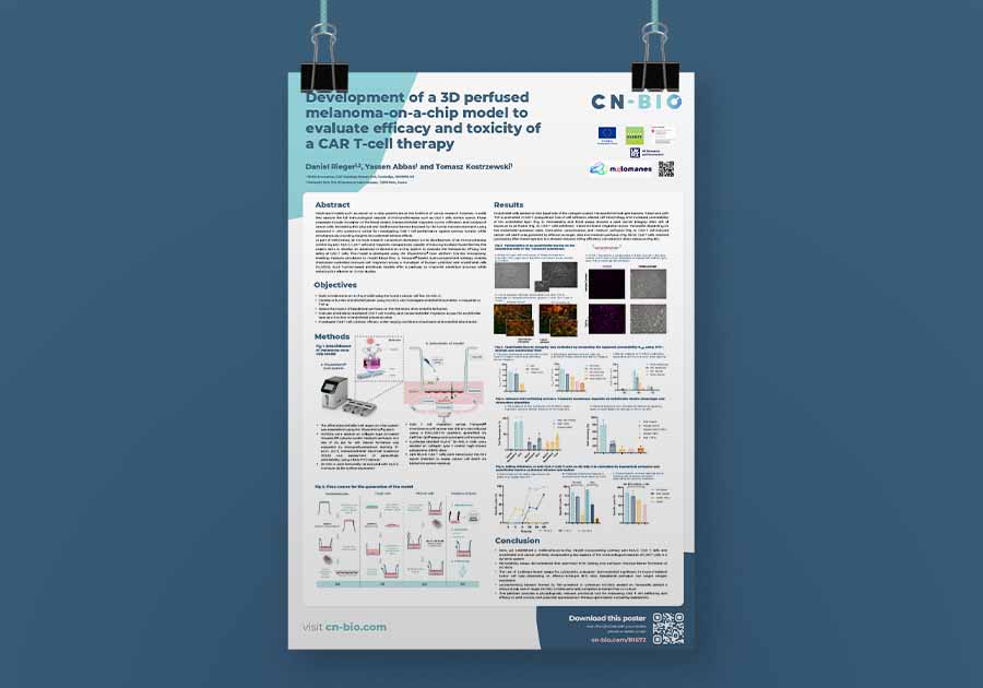
See the full dataset behind the model
The poster sets out the barrier characterization, transmigration, and cell killing data in full, including the conditions that changed how many CAR T cells crossed the barrier and how well they killed once they arrived.
Where cancer-on-a-chip models fit
The melanoma-on-a-chip described here is one configuration of a broader approach. A cancer-on-a-chip puts human tumor cells behind a perfused vascular barrier so that trafficking, infiltration, and killing can be measured as separate events rather than inferred from a single endpoint. Two recent papers illustrate the range. Liu and colleagues, writing in Nature Biotechnology, built a vascularized tumor-on-a-chip using human lung tumor explants and tracked CAR T cell trafficking, activation, and killing across a perfused vascular bed. Maulana and colleagues, in Cell Stem Cell, placed breast cancer aggregates behind an endothelial barrier and followed immune cell transmigration and CAR T efficacy. Both incorporate vasculature, which is the advance that makes trafficking measurable at all.
The tumor compartment is the part that can be exchanged. This model uses SK-MEL-5, an immortalized melanoma cell line, which gives control over antigen expression and keeps the assay reproducible. The same chip design can carry patient-derived material, including tumor explants and organoids, trading some of that control for closer fidelity to an individual patient’s tumor. Which configuration earns its place depends on the question being asked, and running them on one platform is how a team finds out.
The PhysioMimix platform can be adopted to run 3D cancer models under flow, so the perfusion, the barrier design, and the trafficking and killing readouts stay constant while the tumor compartment changes. That continuity is what lets a program swap a controlled cell-line model for patient-relevant material without starting the assay over.
Why this matters for solid tumor programs
For a team developing CAR T therapies against solid tumors, the value of a cancer-on-a-chip is physiological relevance with a purpose. The model captured immune cell extravasation, tumor response, and the drop in efficacy that comes with crossing a barrier, which are the behaviors that decide whether a solid tumor program works. Being able to study immune therapeutic responses in a solid tumor context, under flow, is the capability that static and animal models have not delivered together.
The difference from a conventional co-culture comes down to flow. When medium is perfused, the CAR T cells are introduced into circulation and meet shear, an endothelial wall, and a chemokine gradient before they ever touch a tumor cell. A static assay puts effector and target in the same well and reads the outcome. It can tell you a CAR T cell kills. It cannot tell you whether the cell would have reached the tumor, how the vessel wall changed its numbers, or how much potency it lost in transit.
There are still no FDA-approved CAR T therapies for solid tumors, so the developers who solve the trafficking and efficacy problem stand to open a large space for patients and industry. The model also opens the safety side of the question. The melanoma-on-a-chip work frames the platform as a way to evaluate both efficacy and toxicity and to surface potential adverse effects of a CAR T therapy, so efficacy and safety can be probed in one system rather than in two disconnected assays.
None of this replaces animal studies or clinical work, and it is not meant to. No single model captures the full complexity of human cancer, and human in vitro systems such as cancer-on-a-chip models, alongside animal models and patient data, together build a more predictive picture than any one of them alone. The regulatory ground is shifting the same way, with the US FDA beginning to prioritize new approach methodologies over animal data, which gives human-based cancer models a clearer path into decision-making.
Complexity you can dial up or down
The obvious objection to a model like this is that it is complicated, and that complicated models are slow. Both concerns are fair, and both are addressable. The system is modular. Barrier optimization can be run on its own, transmigration can be measured without the killing step, and the full cascade only has to be assembled when the question needs it. Complexity gets added where it changes the answer and left out where it does not, which is the trade-off every cancer model has to make well: as complex as the biology demands, and no more.
Get started with cancer-on-a-chip models on the PhysioMimix MPS
A perfused melanoma-on-a-chip gives CAR T developers a way to observe the entirety of a therapeutic journey and to see where efficacy is won or lost, and the same platform that runs the cancer-on-a-chip models can also run the patient-derived models that take the approach further. The model described here is an early-stage model built on a human cell line rather than a finished predictive model. What it offers though is a physiologically relevant preclinical tool for studying CAR T cell trafficking and efficacy in solid tumors, with clear applications in therapy optimization and safety assessment.